Category Archives: Imaging

Can I Take A Hypotensive Patient to CT? Part 2

In my last post, I commented on a paper that tried to claim that there is no reason not to take a patient to CT if they are hypotensive. It had issues, as you saw. Today, I want to share another paper from a few years ago that tried to do the same. Again, read the abstract!

I’ve said it before: hypotension and CT scanners don’t play together well. For years I’ve cautioned against this, having seen a number of patients crash and burn in this area early in my career. But it’s a common error, and may jeopardize your patient’s safety. A paper that is now in press looked at this practice in a trauma hospital in Taiwan.

Patients who had blunt abdominal trauma were retrospectively reviewed. Those who remained hypotensive (SBP<90) after 2L of crystalloid were scruitnized. The CT scanner was described as being located in the same area as the ED resuscitation rooms. Furthermore, several physicians and nurses were present during scans, and a full selection of resuscitation equipment was available in the scan area.

Here are the factoids:

  • 909 patients were entered into the study
  • Only 91 patients remained hypotensive after initial resuscitation, and only 58 of these were scanned before definitive management
  • As expected, patients who were hypotensive after initial resuscitation had more serious injuries (ISS 22 vs 12), required more blood transfusions (938 vs 202 cc), and had a higher mortality (10% vs 1%).
  • There were no significant differences in comparing hypotensive patients who went to CT scan vs those who did not if they underwent some sort of hemostatic procedure (laparotomy, angioembolization)
  • In the hypotensive patients, time to OR in the CT scan group was 58 minutes vs 62 minutes for those who skipped the scan.
  • In the same patients, time to angio in the CT scan group was 147 minutes vs 140 minutes without a scan first.

The authors conclude that “hypotension does not always make performing a CT scan unfeasible.” (weak!)

Read this paper closely and don’t get fooled! It is very retrospective and very small. And if you look at the times carefully, you will see some funny business. How can time to OR or angio be virtually identical regardless of whether CT is used? Is it the world’s closest, fastest scanner? Probably not.

The authors showed that hypotensive patients have a ten-fold increase in mortality. They also recognized that definitive control of hemorrhage is the key to saving the patients. Unfortunately, there are factors in this retrospective study, such as various biases and some undocumented factors that make their two patient groups look artificially alike. This gives the appearance that the CT scan makes no difference.

In reality, the fact that there is no difference in times ensures that there is no clinical difference in outcome. To really answer this question, this kind of study must be done prospectively, and must have an adequate population size.

Bottom line: Don’t even consider going to CT with hypotensive patients. Even if you have the fastest, closest scanner in the world. Shock time still kills, and most CT scan rooms are very poor resuscitation rooms. If your patient is unstable in the ED, do your ABCs, get a quick exam, then transport to the area where you can get control of the bleeding. This will nearly always be your OR.

Reference: Hypotension does not always make computed tomography scans unfeasible in the management of blunt trauma patients. Injury, 46(1):29-34, 2015.

The Pan-Scan For Trauma

Diagnostic imaging is a mainstay in diagnosing injuries in major trauma patients. But the big questions are, how much is enough and how much is too much? X-radiation is invisible but not innocuous. Trauma professionals tend to pay little attention to radiation that they can’t see in order to diagnose things they can’t otherwise see. And which may not even be there.

There are two major camps working in emergency departments: scan selectively vs scan everything. It all boils down to a balance between irradiating enough to be satisfied that nothing has been missed, and irradiating too much and causing harm later.

A very enlightening study was published last year from the group at the University of New South Wales. They prospectively looked at their experience while moving from selective scanning to pan-scanning.They studied over 600 patients in each cohort, looking at radiation exposure, missed injuries, and patient injury and discharge disposition variables.

Here are the factoids:

  • Absolute risk of receiving a higher radiation dose increased with pan-scanning from 12% to 20%. This translates to 1 extra person of every 13 evaluated receiving a higher dose.
  • The incidence of receiving >20 mSv radiation dose nearly doubled after pan-scanning. This is the threshold at which we believe that cancer risk changes from low (<1:1000) to moderate (>1:1000).
  • The risk of receiving >20 mSv was lower in less severely injured patients (sigh of relief)
  • There were 6 missed injuries with selective scanning and 4 with pan-scanning (not significant). All were relatively minor.

Bottom line: Granted, the study groups are relatively small, and the science behind radiation risk is not very exact. But this study is very provocative because it shows that radiation dose increases significantly when pan-scan is used, but there was no benefit in terms of decreased missed injury. If we look at the likelihood of being helped vs harmed, patients are 26 times more likely to be harmed in the long term as they are to be helped in the short term. The defensive medicine naysayers will always argue about “that one catastrophic case” that will be missed, but I’m concerned that we’re creating some problems for our patients in the distant future that we are not worrying enough about right now.

Related posts:

Reference: Comparison of radiation exposure of trauma patients from diagnostic radiology procedures before and after the introduction of a panscan protocol. Emerg Med Australasia 24(1):43-51, 2012.

Routine CT After Operative Exploration For Penetrating Trauma

CT scans are commonly used to aid the workup of patients with blunt trauma. They are occasionally useful in penetrating trauma, specifically when penetration into a body cavity is uncertain and the patient has no hard signs that would send him or her immediately to the operating room.

Is there any role in operative penetrating trauma, after the patient has already been to the OR? The dogma has always been that the eyeballs of the surgeon in the OR are better than any other imaging modality. Really?

The surgical group at San Francisco General addressed this question by retrospectively reviewing 6 years of their operative penetrating injury registry data. They were interested in finding how many occult injuries (seen with CT but not by the surgeon) were found on a postop CT. A total of 225 patients who underwent operative management of penetrating abdomen or chest injury were included.

Here are the factoids:

  • Only 110 patients had a postop CT scan; 73 had scans within the first 24 hours, the other 37 were scanned later
  • Rationale for early scan was to investigate retroperitoneal injury in half of patients, but frequently no indication was given (41%)
  • Rationale for late scan was for workup of ileus in one third, or for evaluation of new or unexpected clinical problems
  • Occult injuries were found in about half of early CT patients (52%), and 22% of late CT patients
  • The most common occult injuries were fractures, GU issues, regraded solid organ injury, and unrecognized vascular injuries
  • Several management changes occurred, including

Bottom line: There appears to be a significant benefit to sending some penetrating injury patients to CT in the early postop period. Specifically, those with injury to the retroperitoneum, deep into the liver, near the spine, or with multiple and complicated injuries would benefit. Simple stabs and gunshots that stay away from these areas/structures probably do not need followup imaging. 

Rreference: Routine computed tomography after recent operative exploration for penetrating trauma: What injuries do we miss? J Trauma, published ahead of print, May 11, 2017.

Chest CT vs Chest X-Ray After Chest Tube Insertion

Two days ago, I discussed getting the traditional chest x-ray routinely after chest tube insertion. The answer was yes, it is important even if it appears to be functioning correctly. But yesterday, I also showed you how the chest x-ray can lie.

Remember this image?

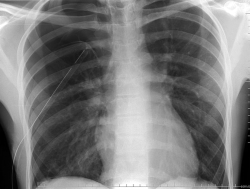
Looks perfect! But it’s a 2-D view and you don’t know where the tube is in the anterior-posterior axis. It turns out to be in the patient’s subcutaneous tissues of his back, near his scapula!

So what if this is a trauma activation patient and you are getting ready to send your patient for a chest CT shortly? Should you follow the usual dogma and still get a conventional chest x-ray prior to leaving the trauma bay?

The answer is no! Typically, your trauma activation patient should have rapid access to the CT scanner, so you won’t have to wait very long. And the additional 3-D information is very helpful in making sure the tube is placed exactly where you want it.

Bottom line: If you are planning on obtaining a chest CT anyway in your trauma patient, don’t bother with a conventional chest x-ray first to check chest tube position. But DON’T order a chest CT for this reason alone! Remember, the chest CT is only for detecting aortic injury in blunt trauma. It should not be used for diagnosing fractures, hemothorax, or pneumothorax. Or chest tube position!

Related posts:


Quiz: Is This A Good Chest Tube?

A blunt trauma activation patient presents with a pneumothorax seen on the initial chest x-ray, obtained in your trauma bay. You professionally insert a large chest tube, and all appears to go well. You shoot a followup chest x-ray and this is what you get:

What do you think of the tube position? Looks great, right?

But if you look carefully, you can see the lung outline in the middle of the right side of the chest. Big-time pneumothorax despite what looks like a perfectly placed tube. There are several possible explanations, and many of you sent me your guesses:

  • The tube is in the lung. This rarely happens to normal lungs. Sure, you can probably do it to an ARDS lung, but otherwise it’s not very likely.
  • The tube is in the fissure. This does happen on occasion, but not often. And many times it works anyway.
  • The tube is occluded or kinked. A PA or AP chest x-ray will show the kink, although bent tubes frequently work anyway. If a hemothorax is present, it is possible that a clot is plugging the tube. Clearing a plugged tube will be the subject of another post.
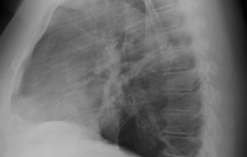
  • It’s not really a chest tube. Hopefully, this would have been detected when it was placed, but it isn’t always. The chest x-ray above looks great, right? Unfortunately, it’s a 2 dimensional representation of a 3-D object. Where is that tube in the z-axis?

In this case the correct answer is the last one. This is one time when I would actually recommend a lateral chest x-ray. Have a look at the result. You can clearly see the tube snaking around into the soft tissues of the back.

Bottom line: Remember that a perfect x-ray doesn’t necessarily mean a perfect tube. Go through the various possibilities quickly, and make it work.

Related posts: