Category Archives: General

Yet Another Paper on Treating Pneumothorax With Oxygen?

Back in February, I thought I closed the door on using high inspired oxygen to try to speed up the resolution of pneumothorax (see related post below). I’ve just run across another attempt which is equally as bad!

This article was published in the Journal of Pediatric Surgery in 2000. The authors randomly divided 27 rabbits into three groups: room air, 40% O2, and 60% O2. Each was given a complete pneumothorax and received chest xrays twice a day. The average time to resolution was measured in each group.

At first glance, it appears that the higher O2 groups resolve faster. But wait, something’s fishy here! In the room air group, the complete pneumothorax went away on its own in 5 days. This doesn’t really happen in people. And in the 60% group, it disappeared in a day and a half! Miraculous!

Oh, and incidentally, a quarter of the rabbits died before completion of the study.

Bottom line: At first glance, these results sure look promising. However, they are rabbits, and they don’t act like people, let alone children! And the resolution times are unrealistic for humans. I still do not recommend the use of high inspired oxygen in an attempt to resolve a pneumothorax. Either some kind of tube is needed for larger volumes (small caliber if air only, bigger if blood is present), or it will go away on its own if the pneumothorax is small.

Related posts:

Reference: Zierold et al. Supplemental oxygen improves resolution of injury-induced pneumothorax. J Pediatric Surg 35(6):998-1001, 2000.

Print Friendly, PDF & Email

Radiation Exposure in Pediatric Trauma

The use of radiographic imaging in trauma patients has exploded over the past decade. A growing amount of research is looking at adult patients, but what about children?

Johns Hopkins did a one year retrospective review of radiographic imaging in kids age 14 and below. The studies performed and the estimated radiation dose was calculated for each child. A total of 719 children were studied and they underwent a total of 4603 studies:

  • CT scans – 1457 (32%)
  • Plain radiographs – 3097 (67%)
  • Fluoroscopy – 49 (1%)

CT accounted for only 32% of studies but delivered 91% of the total radiation dose. Children involved in car crashes received the highest dose of radiation (18mSv) versus burned children, who had the lowest dose (1.2 mSv). Radiation exposure increased as the injury severity increased. The average age was 8 years, which means that these children have a long time until possible side-effects emerge.

What to do? First, seriously weigh the risks and benefits of every radiographic study before you order it. If CT is not essential, do something else. The ALARA concept is key (as low as reasonably achievable):

  • Use weight-based CT protocols in order to deliver the minimum amount of radiation needed to get decent images
  • Shield all sensitive areas that are not being imaged
  • Use focused studies
  • Avoid repeat exams
  • Become knowledgeable about the effects of radiation exposure
  • Ask yourself: “What if this were my child?

Reference: Brown, et al. Diagnostic radiation exposure in pediatric trauma patients. J Trauma 2010, ahead of print. 

Print Friendly, PDF & Email

How Big Was The Knife?

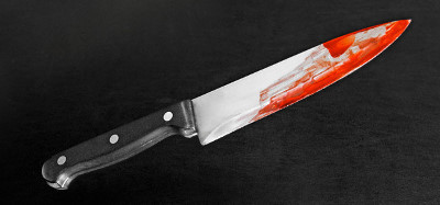
As part of a thorough history and physical on any trauma patient, we typically ask “How big was the knife you were stabbed with?” and “How deep did it go?”

Unfortunately, the answers you typically will get are “This big!” while they hold their hands at least 3 feet apart, and “All the way, doc!”

These answers are not very helpful, so it is not really of much use to ask the questions. The “how big” question is not helpful at all, because a long knife may barely penetrate, and a paring knife can make it all the way into the heart.

This leaves the “how deep” question. There are two ways to determine the answer. If the paramedics or police bring the weapon in, you can carefully examine it (taking proper care to preserve forensic evidence) and see if there is a blood line extending from the tip. This will show the maximum depth of penetration.

The second way is to examine the wound, either by local wound exploration or using CT.

Helpful hint: You can’t tell how large (wide) the blade is by looking at the wound. The elastic nature of the skin causes it to stretch as it is being cut during the stab. When the knife is removed, the resulting laceration will always be less wide than the blade.

Print Friendly, PDF & Email

Making Aeromedical Flights Safer

There are about 840 EMS helicopters operating nationwide. The fatal accident rate has doubled from the mid-90’s to the growth spurt seen in the earlier part of this decade. Since late 2007, 57 crew members and patients have died in these helicopter crashes. According to the FAA, the most frequent causes of these crashes were controlled flight into terrain, inadvertent flight into instrument conditions, and disorientation during night flight.

The FAA is now proposing to change the rules and add extra equipment to these flights in an attempt to improve safety. These changes would include:

  • Installing a ground proximity warning system
  • Tightening the restrictions that limit proximity to bad weather. Currently, pilots must stay half a mile away from clouds during the daytime and one mile away at night.
  • Boosting bad weather training requirements for pilots so they are better equipped to escape from bad weather
  • Installing flight data recorders. New, lightweight models need to be developed for helicopters first, though.

It looks like this is a win-win proposition. Lawmakers, families of crash victims and the aeromedical industry appear to be on board with these changes. Once approved, they would go into effect next year. Unfortunately for the families of crew and patients killed in these crashes, the changes can’t come soon enough.

Print Friendly, PDF & Email

Diagnosing Facial Fractures With CT

Facial fractures are common after major blunt trauma. There are a number of diagnostic tests available for their diagnosis, including head CT, conventional facial imaging and facial CT.

Our preference has been to add a facial CT to the list of diagnostics in any patient with external evidence of facial trauma. Subjectively, it appeared that there were not many injuries being identified, and the vast majority did not require operative management. 

A review of the literature shows that head CT alone is sufficient for screening for significant facial fractures. A small retrospective series noted that the accuracy was 92%, with 90% sensitivity and 95% specificity. 

Bottom line: A head CT alone ordered for the usual indications is a very good screening test for facial fractures. If none are seen, it is unlikely that there are any fractures that require specific management. If fractures are seen, consultation with a facial surgeon is needed. However, unless the fractures involve critical areas (e.g. temporal bone near the middle ear) or are significantly displaced, the benefit of a facial CT scan is still very low since most will be treated without operation.

Reference: Computed tomography of the head as a screening examination for facial fractures. Marinaro et al. Am J Emerg Med 25, 616-619, 2007.

Print Friendly, PDF & Email