Category Archives: General

DVT In Children: How Old Is Old Enough?

Adult trauma patients are at risk for venous thromboembolism (VTE). Children seem not to be. The big question is, when do children become adults? Or, at what age do we need to think about screening and providing prophylaxis to kids? As of yet, there are no national guidelines for dealing with DVT in children.

Researchers at Johns Hopkins went to the NTDB to try to answer this question. They looked at the records of over 400,000 trauma patients aged 21 or less who were admitted to the hospital. 

Here are the interesting factoids:

  • Only 1,655 patients (0.4%) had VTE (1,249 DVT, 332 PE, 74 DVT+PE)
  • VTE patients were older, male, and frequently obese
  • VTE patients were more severely injured, with higher ISS and lower GCS
  • Patients with VTE were more likely to be intubated and receive blood transfusions, and had longer hospital and ICU stays

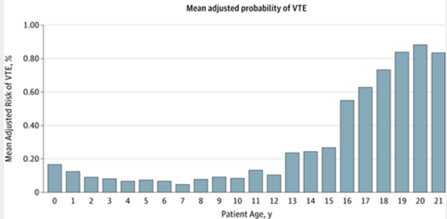
The risk of VTE stratified by age was as follows:

Bottom line: Risk of VTE in pediatric trauma patients follows the usual injury severity pattern. But it also demonstrates a predictable age distribution. Risk increases as the teen years begin (13), and rapidly becomes adult-like at age 16. Begin your standard surveillance practices on all 16 year olds, and consider it in 13+ year olds if their injury severity warrants.

Related post:

Reference: Venous thromboembolism after trauma: when do children become adults. JAMA Surgery online first October 31, 2013.

Hypothermia For Treatment of Severe TBI?

We’ve been trying to figure out therapeutic hypothermia for a long time. Although we know that accidental hypothermia, especially in trauma patients, is not a good thing, it seems to be protective in certain circumstances. The most significant areas of interest center around the neuroprotective effects, especially after ischemia or hypoxia.

But with the good always comes the bad. Every intervention has side effects, and hypothermia is no exception. Decreased cardiac efficiency, blood viscosity increases, pulmonary dysfunction or edema, coagulopathy, decreased tissue oxygen availability, and changes in drug pharmacodynamics are but a few of the problems that may arise. But as long as the benefits outweigh the risks, such an intervention may be acceptable.

We’ve been looking at the possible protective effects of hypothermia on the brain after severe head injury for quite some time. As with most neurotrauma studies, hypothermia ones are tough to do well. Patient selection, adequate numbers of subjects and good randomization and/or blinding are very difficult. It requires assembling all the relevant studies and scrutinizing this whole body of work to figure out if it works or not.

And the answer is, it doesn’t. The Cochrane Library updated their previous work in this area in 2009. They combined 23 studies and over 1600 patients to try to determine if hypothermia (35C for at least 12 hours) is protective in patients with severe TBI. After whittling the field down to good quality studies, they found that there may be a trend toward fewer unfavorable outcomes (death, severe disability, vegetative state), but it was not statistically significant. There were variable results with respect to the incidence of pneumonia after hypothermia, and these, too, did not meet statistical significance.

Bottom line: Therapeutic hypothermia for treatment of severe TBI is still not ready for prime time, and may never be. The studies thus far are small and flawed. Don’t implement your own protocol for this technique unless you are involved in a very high quality, multi-center study that will add to the literature!

Reference: Hypothermia for traumatic head injury. The Cochrane Library 2009, Issue 4.

Dysphagia and Cervical Spine Injury

Cervical spine injury presents a host of problems, but one of the least appreciated ones is dysphagia. Many clinicians don’t even think of it, but it is a relatively common problem, especially in the elderly. Swallowing difficulties may arise for several reasons:

  • Prevertebral soft tissue swelling may occur with high cervical spine injuries, leading to changes in the architecture of the posterior pharynx
  • Rigid cervical collars, such as the Miami J and Aspen, and halo vests all force the neck into a neutral position. Elderly patients may have a natural kyphosis, and this change in positioning may interfere with swallowing. Try extending your neck by about 30 degrees and see how much more difficult it is to swallow.
  • Patients with cervical fractures more commonly need a tracheostomy for ventilatory support and/or have a head injury, and these are well known culprits in dysphagia

A study in the Jan 2011 Journal of Trauma outlines the dysphagia problem seen with placement of a halo vest. They studied a series of 79 of their patients who were treated with a halo. A full 66% had problems with their swallowing evaluation. This problem was associated with a significantly longer ICU stay and a somewhat longer overall hospital stay.

Bottom line: Suspect dysphagia in all patients with cervical fractures, especially the elderly. Carry out a formal swallowing evaluation, and adjust the collar or halo if appropriate. 

Reference: Swallowing dysfunction in trauma patients with cervical spine fractures treated with halo-vest fixation. J Trauma 70(1):46-50, 2011.

Using Mechanism of Injury In Your Trauma Activation Criteria

The Centers for Disease Control and Prevention (CDC) published a set of Guidelines for Field Triage two years ago. Click here to download them. They list 4 tiers of activation criteria to help prehospital providers triage patients appropriately to trauma centers. 

Tier 1, which are physiologic criteria, and Tier 2 (anatomic criteria) are very accurate in predicting injury serious enough to require trauma team activation. Tier 3 contains mechanism criteria, and many centers who use these verbatim in their activation criteria end up with a fair amount of overtriage. Some centers even see a significant number of patients who meet Tier 3 criteria go home from the ED!

The Yale department of Emergency Medicine looked at intrusion into vehicle criteria (more than 12" near an occupant, more than 18" anywhere on the vehicle) to see if they are a valid predictor for admission or trauma center transport. It was a retrospective review of EMS transports to the Yale ED or to one satellite site. 

Unfortunately, the number of vehicles that met intrusion criteria (48) was small compared to the number without significant intrusion (560). This makes the data a little less convincing than it may have been. The likelihood that intrusion would require trauma center admission (Positive Predictive Value) was only 26%. The likelihood that trauma center resources would be utilized (for issues like death, ICU stay, operation, spinal injury or intracranial hemorrhage) was only 13%. The authors recommend that the CDC guidelines be tweaked based on this data.

Bottom line: I think the numbers are far too small to convince the CDC to change their guidelines. But I would urge each trauma center that uses the intrusion criteria for activation to carefully study how many of those patients have minor injuries or go home from the emergency department. They may find that they can rely on other more accurate criteria and decrease their overtriage rate at the same time.

Reference: Motor vehicle intrusion alone does not predict trauma center admission or use of trauma center resources. Prehospital Emerg Care 15:203-207, 2011.

The Tripod Fracture

The tripod fracture (officially known as the zygomaticomaxillary complex fracture, and sometimes called a malar fracture) is the most common one seen after trauma. Fundamentally, the zygoma is separated from the rest of the face in a tripod fracture.

As you might imagine (tripod fracture), there are three components to this fracture. The first is a fracture through the zygomatic arch (1). Next, the fracture extends across the floor of the orbit and includes the maxillary sinus (2). Finally, the fracture includes the lateral orbital rim and wall (3). 

Extraocular muscles may become trapped in the fracture line, leading to diplopia. It is very important to do a good eye exam to try to detect entrapment. The infraorbital nerve also passes through the orbital floor and may be injured, leading to numbness along the lower eyelid and upper lip.

Nondisplaced fractures are treated symptomatically and reevaluated after a week or so to see if surgery would be beneficial. Displaced or symptomatic fractures require early open reduction. The pictures below show the anatomy of these fractures. They are derived from teaching materials provided by the Radiology Department at the University of Washington.

image

image