Category Archives: Complications

NSAIDs And Fracture Healing Revisited – Yet Again!

I’ve written so many posts about the use of non-steroidal anti-inflammatory drugs (NSAIDs) it’s practically getting old. To summarize, some old animal studies suggested that using NSAIDs during fracture healing could impair the process. However, human studies were not so convincing.

Over the years, there has been quite a bit of conflicting evidence. This generally means the association between healing and NSAID use is weak. However, after this period of time, we should have become aware of a significant cause/effect relationship.

The Eastern Association for the Surgery of Trauma recently released a practice management guideline regarding the use of NSAIDs for the treatment of acute pain after orthopedic trauma. They used a standard methodology to identify and analyze published research. They focused on human studies specifically relating to this drug class’s use in fractures. The group ultimately identified 19 pertinent research papers for analysis, 10 of which were prospective, randomized studies.

Here are the three questions they asked, with their answers:

  • Should NSAIDs be used in analgesic regimens for adult patients
    (≥18 years old) with traumatic fracture versus routine analgesic
    regimens that do not include NSAIDs to improve analgesia and
    reduce opioid use without increases in non-union and acute kidney
    injury rates? Although the quality of the studies for this question was low, EAST conditionally recommended using NSAIDs in pain control regimens. In the higher-quality studies in this group, there was no increased risk of non-union.
  • Should ketorolac be used in analgesic regimens for adult patients with traumatic fracture versus routine analgesic regimens that do not include ketorolac to improve analgesia and reduce opioid use without increasing non-union
    rates? This is the same question asked above, but with a specific drug rather than the class in general. The answer was basically the same.
  • Should selective NSAIDs (COX-2 inhibitors) be used in analgesic
    regimens for adult patients (≥18 years old) with traumatic fracture versus routine analgesic regimens that include non-selective NSAIDs to improve analgesia and reduce opioid use without increasing non-union rates?
    COX-2 inhibitors are a subset of NSAIDs that are more selective in their action, blocking only the COX-2 receptor. Several years ago, there was a scandal regarding the COX-2 inhibitor rofecoxib (Vioxx). These selective drugs tended to have a higher incidence of cardiac complications. The manufacturer covered up this fact for several years, resulting in many unneeded deaths before it was removed from the market. The only COX-2 inhibitor available in the US is celecoxib. Only a few studies were performed using this drug during bone healing. There were not enough to make a recommendation.

Bottom line: EAST made conditional recommendations for using NSAIDs in general and ketorolac specifically in adults with fractures. “Conditional” only means that the authors did not have a consensus. Some voted to strongly recommend, and the remainder to conditionally recommend. There were no votes to recommend against their use.

The use of NSAIDs should complement a well-thought-out opioid regimen, which should also be combined with other non-narcotic medications and appropriate mobilization and therapy.

Reference: Efficacy and safety of non-steroidal anti-inflammatory
drugs (NSAIDs) for the treatment of acute pain after orthopedic trauma: a practice management guideline from the Eastern Association for the Surgery of Trauma and the Orthopedic Trauma Association. Trauma Surg Acute Care Open. 2023 Feb 21;8(1):e001056. doi: 10.1136/tsaco-2022-001056. PMID: 36844371; PMCID: PMC9945020.

Top 10 Worst Complications: #1 Nasocerebral Tube

Minor complications from nasogastric tube insertion occur relatively frequently. Emesis is fairly common when the gag reflex is stimulated by the tube in the back of the oropharynx. An infrequent but possibly fatal one is insertion through the cribriform plate. 

The cribriform plate is located directly posterior to the nares and is part of the ethmoid bone. It is very porous in nature and weaker than the surrounding portions of the ethmoid. It is easily fractured, and can be seen is association with basilar skull fractures. This is one source for rhinorrhea in patients with these fractures.

Cribriform fracture is a contraindication to unprotected insertion of a nasogastric tube. If you look at the sagittal section below, the plate lies directly behind the nares. When inserting the NG tube, we are usually taught to aim the tube straight back. Unfortunately, this aims it directly at the cribriform. If a fracture is present, it is possible that you may be inserting a nasocerebral tube!

Cribriform plate - sagittal section

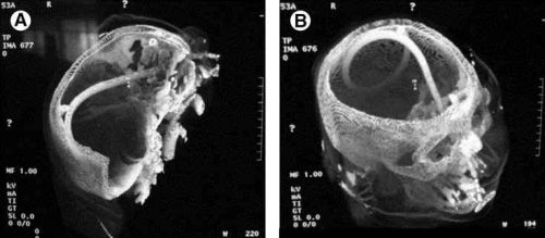
The usual symptoms when this occurs consist of immediate neurologic deterioration to coma, and a unilateral or bilateral blown pupil. The tube must not be withdrawn, because it will cause significant injury to the base of the brain. A stat neurosurgical consultation must be obtained, and if the patient is salvageable, the tube must be withdrawn through a craniectomy.

To avoid this dreaded complication, identify patients at risk for cribriform injury. They are:

  • patients with signs of trauma from eyebrows to zygoma
  • comatose patients
  • patients with signs of basilar skull fracture (Battle’s sign, raccoon eyes, oto- or rhinorrhea)

If your patient is at risk, follow these guidelines:

  • first, does the patient really need a gastric tube?
  • if comatose, insert an orogastric tube
  • if awake, don’t put the tube in their mouth, as they will gag continuously. Instead, place a lubricated, curved nasal airway. Then lube up a slightly smaller Salem sump tube and pass it through the airway.

Nausea In The Trauma Bay: Gastric Tube vs Anti-Emetic Drugs?

Nausea and vomiting are common problems in trauma patients, particularly those in a trauma activation. Inciting factors include pain, full stomach from food eaten before the event or blood swallowed after, or reaction to pain medications. For years, trauma professionals reached for the lowly gastric tube to evacuate stomach contents to “solve” the problem.

But how many of you have seen a patient forcefully empty their stomach as soon as the tube touches the oropharynx? And of course, your patient is lying supine, so the vomitus goes straight up, then back down into their airway. And if their mental status is not quite right, they may aspirate, causing even bigger problems.

We’ve had anti-emetic medications for a long time, some more effective than others. Only recently have we begun to rely on these as a first line defense in the trauma resuscitation room. But do they work? Are they safer?

The University Medical Center Utrecht in the Netherlands looked at this problem. They changed their policy from inserting a gastric tube to administering anti-emetics at the beginning of 2014. They studied their experience for the 6 months before and 6 months after the policy change. They inserted an orogastric (OG) tube preferentially before the switch, and used ondansetron and/or metoclopramide after.

Here are the factoids:

  • A total of 1446 trauma patients were admitted during this period. After excluding patients who were intubated or did not complain of nausea, 453 were analyzed (30%)
  • 20% of patients who had an OG tube placed vomited vs only 3% receiving medication (significant)
  • After therapy, 14% of patients receiving an OG were still nauseated vs only 2% getting meds (also significant)
  • 3 patients vomited and aspirated after OG placement, and 1 developed a pneumonia. 2 patients became bradycardic and med administration, and one developed QT-prolongation

Bottom line: This is a relatively small, retrospective study. Furthermore, the choice of gastric tube route (oral) is a setup for gagging and vomiting. Nasogastric tubes are a bit less noxious, but can’t be inserted in all patients (see next week’s post). Even so, the use of anti-emetics in trauma patients complaining of nausea seems like the kinder, gentler way to go. 

Which drug to use? Previous studies have shown that ondansetron 4mg is as effective as 8mg, and that this drug is about equally as effective as metoclopramide. There is also some evidence that giving both is more effective than just giving one.

Gastric tubes are still important, particularly in the comatose patient. But since these patients are at risk for cribriform plate injury, only the oral route should be used.

Reference: Analysis of two treatment modalities for the prevention of vomiting after trauma: orogastric tube or anti-emetics. Injury (accepted manuscript, in press) online 8 July 2017.

Colonic Pseudo-Obstruction In Trauma Patients – Part 2

In my last post, I discussed a paper describing the incidence of colonic pseudo-obstruction (CPO), or Ogilvie syndrome, in trauma patients. The paper confirmed my bias that this condition could be a problem in a specific subset of trauma patients. They are generally older men with pelvic or spine fractures, with or without surgical fixation. In addition, some comorbidities like diabetes, obesity, and concomitant head injury increase the incidence.

The usual dogma is that a cecal diameter > 12cm places the patient at risk of perforation. Therefore, as the size of the colon increases, steps should be taken to decompress it definitively. This typically involves neostigmine infusion, which usually requires transfer to the ICU, or colonoscopic decompression.

Until about eight years ago, we managed this issue at Regions Hospital using the IV neostigmine option in the ICU. But then, one of our colorectal surgeons described his experience managing CPO with subcutaneous neostigmine. A light bulb turned on! Intravenous neostigmine requires admission to an ICU at our hospital for continuous monitoring to quickly identify the development of bradycardia.

But subcutaneous neostigmine was not on the naughty list! We developed a practice guideline to identify and exclude patients for whom this drug was contraindicated. And it required monitoring that could be accomplished in a floor bed with brief episodes of continuous EKG monitoring. Our inpatient trauma unit could easily do this. However, it might require a step-down bed in yours.

Here is the guideline. Click the image of the link at the end of this post to download a copy.

Here are the major features of the guideline:

  • Identification. Any patient, especially those with the previously described risk factors, begins daily monitoring with a flat plate abdominal x-ray. Patients with abdominal distension with subjective discomfort or nursing concerns with the distension fall into this category.
  • Trigger. Once distension of any part of the colon, particularly the cecum, exceeds 10 cm, it is time to act. Otherwise, daily monitoring and a bowel regimen continue.
  • Contraindications to neostigmine. If the patient has a recent history of MI, bronchospasm, is on beta-blocker therapy, or has SBP < 90 torr, heart rate < 60, or weight < 50kg, colonoscopic decompression should be carried out.
  • Continuous monitoring must be available for one hour after injection. This requires an appropriate nurse and an EKG monitor. Atropine must be present at the bedside in case bradycardia develops.
  • Up to three doses of SQ neostigmine (1mg) can be given 12 hours apart. If the patient responds with a large bowel movement or passage of gas, it should be confirmed with an abdominal x-ray.
  • Patients with insufficient response must transfer to ICU for IV neostigmine or should be scheduled for an urgent colonoscopy.

Our experience has shown that this guideline is usually very effective. However, a few patients have had a recurrence after 24-48 hours, which is uncommon. The guideline can be repeated if necessary.

Bottom line: A low index of suspicion for CPO in trauma patients is critical. Once the colon perforates, these patients do poorly, and serious complications are common. This guideline allows the trauma service to keep these patients out of the ICU while treating it. But before you implement this, please work closely with your pharmacists to ensure that hospital policy allows using neostigmine outside of an ICU setting.

Colonic Pseudo-Obstruction in Trauma – Practice guideline. Click to download.

Colonic Pseudo-Obstruction In Trauma Patients – Part I

A funny thing happened eight years ago. During one of our morbidity and mortality conferences at Regions Hospital, we got the first hint of an emerging pattern. We noted occasional trauma patients who developed colonic pseudo-obstruction (CPO), also known as Ogilvie’s syndrome.

In reviewing our experience, it seemed to occur mostly in men who had sustained pelvic or thoracolumbar spine injuries. Surgical instrumentation for these injuries also appeared to be a common factor, as was middle-aged or older, obesity, and metabolic diseases like type II diabetes.

We continued to see the pattern and treated it in a highly variable way depending on the attending surgeon. Abdominal x-rays were obtained semi-randomly, and if the cecum was considered as the ill-defined term “large,” the patient was sent to the ICU for an injection of neostigmine or endoscopic evacuation. If a perforation occurred, patients often got very sick.

As always, variable practice patterns are fodder for developing a practice guideline. This is the first part of a two-part series on CPO in trauma patients. First, I’ll review a new article describing this condition’s incidence in orthopedic patients. Then, in my next post, I will share a practice guideline we developed for use at Regions Hospital.

The paper was a retrospective cohort study performed by the surgical group at Copenhagen University Hospital in Denmark. They focused on patients who underwent pelvic or acetabular procedures for traumatic injury over twelve years. One cohort consisted of patients who developed CPO; the other did not.

The definition of CPO was based on standard procedures that this surgical group already used, although the specifics were not fully explained. It was based on a physical examination of the abdomen, laboratory tests, and radiographic images. Patients with a colonic diameter >10 cm were treated with neostigmine infusion. Colonoscopic decompression was used if neostigmine did not work or was contraindicated.

Here are the factoids:

  • Of 1060 patients who underwent pelvic or acetabular procedures for trauma, 25 developed CPO (2.4%)
  • The incidence was only 1.6% for pelvic fractures and about 2.6% for acetabular fractures or combined fracture patterns
  • Risk factors identified included motorcycle crash, preperitoneal packing, concomitant skull fracture or intracranial hemorrhage, paraplegia or tetraplegia, internal fixation, congestive heart failure, diabetes, and sepsis or nosocomial infection
  • CPO development increased ICU length of stay by 9 days and added a month to the hospital stay
  • Mortality was higher in the CPO group (8% vs. 6%), but this was not statistically significant

Bottom line: This is the first paper I’m aware of that quantifies what I have already seen regarding Ogilvie’s syndrome in trauma. It should be an eye-opener for everyone who sees seriously injured orthopedic patients. The increased lengths of stay are enormous, which adds to the cost and the potential for even more complications.

Obviously, this is a problem that needs to be taken very seriously. Use of the ICU for neostigmine infusion or procedural decompression should be common. But recognition and initial management should be standardized, so all appropriate patients are treated for the condition.

In my next post, I’ll share the practice guideline we developed at Regions hospital. It is designed to identify the condition early and provide decompressive therapy without moving the patient to the ICU.

Reference: Ogilvie Syndrome in Patients With Traumatic Pelvic and/or
Acetabular Fractures: A Retrospective Cohort Study. J Orthop Trauma 37(3):122-129, 2023.