Category Archives: Complications

Inserting an NG Tube (Not an NC Tube)!

On occasion (but not routinely) trauma patients need to have their stomach decompressed. The reflex maneuver is to insert a nasogastric (NG) tube. However, this may be a dangerous procedure in some patients.

Some patients may be at risk for a cribriform plate fracture, and blindly passing a tube into their nose may result in a nasocerebral (NC) tube (see picture). Immediate and profound neurologic decompensation usually occurs. This is a neurosurgical catastrophe, and the outcome is uniformly dismal. It generally requires craniectomy to remove the tube.

The following patients are at risk:

  • Evidence of midface trauma (eyebrows to zygoma)
  • Evidence of basilar skull fracture (raccoon eyes, Battle’s sign, fluids leaking from ears or nose)
  • Coma (GCS<8)

If you really need the tube, what can you do? If the patient is comatose, it’s easy: just insert an orogastric (OG) tube. However, that is not an option in awake patients; they will continuously gag on the tube. In that case, lubricate a curved nasal trumpet and gently insert it into the nose. The curve will safely move it past the cribriform plate area. Then lubricate a smaller gastric tube and pass it through the trumpet.

Incidental Appendectomy During Trauma Laparotomy?

The debate over incidental appendectomy has waxed and waned over the years. And for the most part, it has nearly permanently waned in general surgical cases for now. But every once in a while, I am asked about incidental appendectomy during trauma laparotomy. Is it a good idea? What reasons could there possibly be for doing it?

In the old days, we would frequently do an incidental appendectomy because… well, just because we were there. The surgeon was in the midst of a general surgical case, typically an open one, and this normal little appendix was just staring us in the face. The justification was usually, “we’ll save him another operation in the future in case he develops acute appendicitis.”

Legitimate reason? It took many years for the literature to develop, but it finally did. Here were the reasons we figured out not to do it:

  • Despite how innocuous a procedure it seems to be, there is a measurable uptick in complication rates. This is true in the usual clean contaminated general surgery cases. Some papers also noted an increased mortality when the appendectomy was added to a cholecystectomy case. In a trauma procedure with bowel injury and contamination, it’s a bit harder to see the correlation. But any time we cut or staple something out, there is always the possibility that it might break down.
  • Cost increases in laparoscopic cases if additional ports and/or equipment is needed for the appendectomy. This doesn’t really apply to major trauma cases, since we better not be doing them laparoscopically!
  • The appendix is not the useless vestigial structure we originally thought. There is evidence that it is a repository for the gut microbiome, which can help repopulate the colon with bacteria after a serious insult like prolonged antibiotic administration. Unnecessary removal may ultimately interfere with gut health and disease.

Can acute appendicitis develop after trauma laparotomy? Sure, at any time. Thankfully, it’s not very common. The presenting complaints are the same as we learned in the doctor books. However, the location of the pain and tenderness may not be in the classic location depending on the post-trauma anatomy and presence of adhesions.

Bottom line: Incidental appendectomy is no longer indicated for just about anything, including trauma laparotomy. If one of your patients presents with abdominal pain at any time, both post-traumatic and other causes must be considered. CT has become the standard for appendicitis workup, and is extremely helpful in sorting out causes in the post-op trauma patient. Use it, and if it is one of the rare cases where appendicitis is actually present, then proceed with the usual and appropriate operative on nonoperative management.

References:

  • Incidental appendicectomy with laparotomy for trauma. Br J Surg 62(6):487-9, 1975
  • Appendicitis following blunt abdominal trauma. Am J Emerg Med 35(9):1386.e5-1386, 2017.
  • Systematic review of blunt abdominal trauma as a cause of acute appendicitis. Ann R Coll Surg Engl 92(6):477-82, 2010.

More On Lead Poisoning And Retained Bullets

Trauma professionals frequently have to leave bullets in patients. It is often more disruptive to go digging the projectiles out than to just leave them in place. But patients always want to know why and what the consequences might be.

In my last post, I discussed a very old paper on what we know about lead levels and retained bullets. Very recently, a meta-analysis was published that provides a better picture of this topic. They somehow managed to find over 2000 articles dealing with lead toxicity and bullets out there. But after someone had the pleasure of reviewing each of them, they found only 12 that had any meaningful or actionable information.

Here are the factoids:

  • All studies were observational (duh! It would be difficult to get your IRB to approve a study where patients were shot on purpose)
  • There were five cross-sectional studies, four case-control studies, and three prospective cohort studies
  • The studies were small, with a median of only 26 patients (range 15-120)
  • Eleven of the twelve studies showed an association with retained bullets and elevated blood lead levels
  • Three studies showed elevated blood levels if a fracture was present
  • The higher the number of retained fragments, the more likely lead levels were to be high
  • Higher lead levels were associated with retained fragments near a bone or joint
  • There were no good correlations with number of fragments and location vs actual lead toxicity

Bottom line: Even using meta-analysis, it is difficult to tease out meaningful answers to this question. That speaks to the low numbers of papers and their quality. However, this study does provide a little bit of guidance.

Retained bullet fragments are probably not a big worry in most patients. The bothersome cases are those where the fragments are in or near a bone or joint. And even though few patients actually developed lead toxicity, lead levels approaching 5 micrograms/dL can have physiologically significant negative effects. 

Recommendation: If your patient has a retained bullet fragment near a bone or joint, or they have “multiple” retained fragments (no good definition of this), they should have blood lead levels measured every three months for a year. If the level is rising, and certainly if it reaches the 5μ/dL level, attempts should be made to remove the fragments.

Reference: Lead toxicity from retained bullet fragments: A systematic review and meta-analysis. J Trauma 87(3):707-716, 2019.

Can Lead Poisoning Occur After A Gunshot?

This is a fairly common question from victims of gunshots and their families. As you know, bullets are routinely left in place unless they are superficial. It may cause more damage to try to extract one, especially if it has come to rest in a deep location. But is there danger in leaving the bullet alone?

One of the classic papers on this topic was published in 1982 by Erwin Thal at Parkland Hospital in Dallas. The paper recounted a series of 16 patients who had developed signs and symptoms of lead poisoning (plumbism) after a gunshot or shotgun injury. The common thread in these cases was that the injury involved a joint or bursa near a joint. In some cases the missile passed through the joint/bursa but came to rest nearby, and a synovial pseudocyst formed which included the piece of lead. The joint fluid bathing the projectile caused lead to leach into the circulation.

The patients in the Parkland paper developed symptoms anywhere from 3 days to 40 years after injury. As is the case with plumbism, symptoms were variable and nonspecific. Patients presented with abdominal pain, anemia, cognitive problems, renal dysfunction and seizures to name a few.

Bottom line: Any patient with a bullet or lead shot that is located in or near a joint or bursa should have the missile(s) promptly and surgically removed. Any lead that has come to rest within the GI tract (particularly the stomach) must be removed as well. If a patient presents with odd symptoms and has a history of a retained bullet, obtain a toxicology consult and begin a workup for lead poisoning. If levels are elevated, the missile must be extracted. Chelation therapy should be started preop because manipulation of the site may further increase lead levels. The missile and any stained tissues or pseudocyst must be removed in their entirety.

Granted, this is a very old paper. Over the years, a few papers on the topic have popped up from time to time. In my next post, I’ll review a meta-analysis on this topic that was just recently published.

Reference: Lead poisoning from retained bullets. Ann Surg 195(3):305-313, 1982.

AAST 2019 #6: DOACs Part 3!

A little further down the direct oral anticoagulants (DOACs) rabbit hole please? The abstract reviewed in my last post suggested that elderly patients taking these agents actually do better than those on warfarin. So if that’s the case, do we need to be so attentive to getting followup CT scans on these patients to ensure that nothing new and unexpected is happening?

The trauma group at UCSF – East Bay performed a multi-center review of the experience at “multiple” Level I trauma centers over a three year period. They included anticoagulated patients with blunt trauma who had a negative initial head CT. Patients taking only an anti-platelet agent or a non-oral anticoagulant were excluded.  They analyzed the data for new, delayed intracranial hemorrhage, use of reversal agents, neurosurgical intervention, readmission, and death.

Here are the factoids:

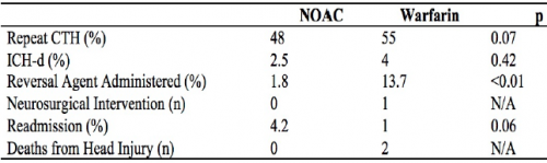
  • A total of 739 records were studied: 409 on warfarin and 330 on a DOAC. Average age was 79, and half were male.
  • Repeat head CT was performed only half the time (!)
  • Delayed hemorrhage was noted in 4% of warfarin cases (9 of 224) and 2.5% of DOAC cases (4 of 159)
  • There were no interventions or deaths in the DOAC group with followup CT, or in those who did not have the repeat scan
  • There was 1 intervention in the warfarin group and two deaths attributed to TBI
  • Reversal agents were administered to 2% of DOAC patients and 14% of warfarin patients
  • The authors performed a regression analysis that showed the two strong associations with delayed hemorrhage were male sex and AIS head > 2 (!)

The authors concluded that this “largest study” suggests that DOACs “may” have a better safety profile compared to warfarin and repeat head CT is not indicated.

Now, hold on a minute!

Rule #1: No single published paper should ever change your practice. They need to be confirmed by other, hopefully better work.

Rule #2: No single abstract should make you even think about changing your practice! These are preliminary works that always need more detail, more effort, and a lot more thought. They are meant to telegraph what the authors are working on and to raise interesting questions from the audience. They should stimulate others to try to replicate and improve upon the work. In general, if something looks really good as an abstract, the next step is successful publication. This means that peers have reviewed the data and agree that it looks promising. But then it should take several years of work by the original authors and others to prove or refute the claims.

This study was small in the first place, and became smaller because half did not have repeat CT scans. The only statistically significant result was that we confirmed that the providers were not very good about getting followup scans. Just because they didn’t do it doesn’t mean it’s not indicated, especially given the nature of the data and the very small numbers.

I consider this another very small piece in the puzzle that suggests DOACs are not as evil as warfarin. There are several of these low power studies floating around right now. But we need to hunker down and really do a big study right so we can start to get a clearer picture of what we should do. For now, it’s best to treat all anticoagulants and anti-platelet agents as evil and err on the side of overtreating.

Here are my comments and questions for the presenter and authors:

  • Why was the followup head CT rate so poor? Was this a “however they like to do it” thing, was there a protocol, did the trauma centers just not believe that DOACs could be bad?
  • What were the guidelines for reversal? If the initial head CT was normal, why ever reverse? This suggests that participating centers could do whatever they wanted based on unspecified criteria.
  • Was the regression analysis helpful in any way? Being male and having a mild TBI seem rather nonspecific factors and wouldn’t help select patients for reversal or repeat scan.
  • Please provide more information on the warfarin intervention and deaths.
  • Isn’t the title of this abstract rather bold for the quality of the results presented?

I’m sure there will be some lively debate at the end of this presentation!

Reference: Repeat CT head scan is not indicated in trauma patients taking novel anticoagulation: a multi-institutional study. AAST 2019, Oral Abstract #66.