How To Manage TBI In Patients On Warfarin

We all know that the combination of traumatic brain injury (TBI) and warfarin can be dangerous. Here at Regions, we developed a reversal protocol a few years ago. However, we found that just having a list of preferred “antidotes” to give was not enough. The time factor is very important, and we found that we needed to ensure prompt use of these medications when indicated.

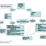
So we added features that ensured timely response and reversal. You can download the protocol by clicking the image above or the link at the bottom of this post.

First, we recognized that any patient with a known or suspected TBI who was taking warfarin was at risk. If the initial GCS was <14, then a full trauma team activation is called. This gives the patient priority lab processing and immediate access to the CT scan. In addition, 2 units of thawed plasma are administered while in the resuscitation room. If the head CT is negative, plasma is stopped.

For patients with a GCS of 14 or 15, a “Code RED” is called, ensuring that an ED physician sees the patient immediately. A point of care INR is drawn and the patient is sent for stat head CT. If the head CT is negative with INR>2.5, the patient is admitted for observation and a repeat head CT is obtained 12 hours later. We have seen patients develop delayed hemorrhage when they have high INR.

We apply a restrictive set of criteria to determine if a patient may go home from the ED, which causes us to admit most for observation. And if they do have a positive CT, we use the algoritm listed below for comprehensive management and reversal.

Bottom line: Patients with any head trauma and an elevated INR are a walking time bomb. They need prompt assessment and reversal of their anticoagulation if indicated. Feel free to share your protocols here as well by posting a comment.

Download the full protocol; click here.

Related post:

Dealing With Shotgun Wounds

Shotgun wounds are uncommon but potentially disabling or deadly. Here are some basics for dealing with them.

Most people only think about the shot that comes out of the shotgun shell and its effects. While this certainly causes a problem, this foreign body is only a part of it. And it can be challenging to pick them out surgically. However, there are two other foreign bodies that become an issue: the wad, and other material on or around the patient (clothing, pieces of a door, etc). 

Look at the diagram above on the left. The wad is a plastic cup that holds the shot and helps the exploding powder charge propel it out of the shell (see sample pictures on the right). If the shotgun is fired at relatively close range, the wad can enter the patient’s wound. It is usually found near the surface and is easily recognized. Be sure to look for it when removing clothing and dressings from your patient.

Any debris from the patient’s surroundings will end up deep to the wad. It is usually centrally located in the wound, and may penetrate as deep as the shot. I once found a piece of coat fabric in the middle of an abscess that formed months after the shooting.

Management of these wounds requires general anesthesia and patience. The soft tissue injury is always worse than it appears, and make take up to 3 days to fully declare itself. I recommend debriding all obviously devitalized tissue and then placing a negative pressure dressing (if possible) to help reduce tissue edema.

All easily found shot should be removed during the initial operation. Deeply embedded particles will either scar in place or migrate to the surface. If all dead tissue is removed, infection is not that common. Inspection of all vital structures, especially blood vessels, should be performed at that time. Remember, due to the velocity and force involved, arteries may appear completely normal on the outside but have significant intimal interruption.

The patient should return to the OR every other day until all tissue die-back has stopped. Final closure or reconstruction can be considered at that time.

Determining The Age Of Bruises

On occasion, you may encounter a patient who has bruising and wonder how old the injuries are. Or there may be several bruises and you would like to know if they occurred at different times. This becomes especially important when dealing with injured children in whom there is a suspicion of abuse.

Bruising occurs when blood leaks from blood vessels into the skin and subcutaneous tissues. If the skin and soft tissues are firm, bruising is not as apparent. In areas where the skin and soft tissues are loose, such as the peri-orbital areas and scrotum, bruising is visible early and may be extensive. The elderly tend to bruise easily because both the skin and subcutaneous tissue are very thin and friable.

A predictable series of color changes occurs with most bruises. During the acute phase, the color is usually reddish and the area may be raised and tender. After about 2 days, the color turns purple and any swelling usually disappears. Over the next week, the color changes to green and yellow as the heme metabolizes. Finally, the color fades and by two weeks most evidence of the injury is gone.

The table above is a key to estimating bruise age. However, this is not an exact science! A number of studies have been performed showing that examiners given photographs of bruises of various ages were not terribly accurate. Fresh and intermediate bruises were identified fairly accurately, but not so for older bruises.

The trauma professional may find it helpful to use these guidelines when trying to decide if there are both fresh and older bruises present. This may indicate that an older adult may be suffering from frequent falls, or that a child needs to be evaluated for abuse.

Reference: Estimation of the age of bruising. Arch Dis Childhood 74:53-55, 1996.

Bucket Handle Injury – Part 2

Yesterday, I wrote about the basics of bucket handle injuries of the intestine. Today, I’ll deal with diagnosing them.

An understanding of the mechanism of injury and a good physical exam are paramount. If the patient took a significant blow to the abdomen, especially in a motor vehicle crash (lap belt), be very suspicious. Any abdominal pain is of concern, particularly in the right lower quadrant (most common injury location). If a CT is indicated and there are focal changes in the mesenery or bowel wall, a trip to the OR is advised.

In some patients, the bowel is devascularized and takes 2-3 days to become necrotic. They experience slowly increasing focal pain, and once this develops it’s time to go to the operating room.

Intubated and/or comatose patients can be problematic in making this diagnosis. There is no physical exam, so the trauma professional has to rely on surrogates. The white blood cell (WBC) count is very helpful. The WBC count is typically elevated into the 15,000-20,000 range immediately after trauma, and declines to normal within about 12 hours. If it begins to climb again after 24 hours, especially if it exceeds 20,000, an intestinal injury is likely.

CT scan and abdominal ultrasound are also helpful. A repeat CT scan may show a change in the volume of fluid, or a change in its character. If the amount of fluid increases significantly, or if a fluid bi-layer is seen, a bucket handle injury is very likely. These findings are pertinent in awake patients as well, but the physical exam usually makes use of these diagnostics unnecessary.

Related posts:

Bucket Handle Injury – Part 1

Bucket Handle Injury

A bucket handle injury is a type of mesenteric injury of the intestine. The intestine itself separates from the mesentery, leaving a devascularized segment of bowel that looks like the handle on a bucket (get it?).

These injuries can occur after blunt trauma to the abdomen. The force required is rather extreme, so the usual mechanism is motor vehicle crash. In theory, it could occur after a fall from a significant height, and I have seen once case where a wood fragment was hurled at the abdomen by a malfunctioning lathe.

The mechanics of this injury are related to fixed vs mobile structures in the abdomen. Injuries tend to occur adjacent to areas of the intestine that are fixed, such as the cecum, ligament of Treitz, colonic flexures and rectum. During sudden deceleration, portions of the intestine adjacent to these areas continue to move, pulling on the nearby attachments. This causes the intestine itself to pull off of its mesentery.

The terminal ileum is the most common site for bucket handle tears. Proximal jejunum, transverse colon, and sigmoid colon are other possible areas. The picture above shows multiple bucket handle injuries in one patient. There are 3 injuries in the small bowel, and one involving the entire transverse colon. Note the obviously devascularized segment at the bottom center of the photo.

Always think about the possibility of this injury in patients with very high speed decelerations. Seat belt marks have a particularly high association with this injury. If your patient has an abnormal exam in the right lower quadrant, or if the CT shows unusual changes there (“dirty” mesenteric fat, thickened bowel wall, extravasation), I recommend a trip to the OR. In these cases, an injury will nearly always be present.

Tomorrow: These injuries can be subtle in an awake patient with a reliable exam. On Friday I’ll write about how you can detect it in unconscious patients.

Source: personal archive. Not treated at Regions Hospital

Home of the Trauma Professional's Blog

Do you want to get a daily email every time there’s a new post? See what I’m up to.

Click here to get details and subscribe!

[accua-form fid=”1″]

[mc4wp_form id=”2023″]