All posts by The Trauma Pro

EAST 2018 #1: Plasma Over-Resuscitation And Mortality In Pediatric TBI

The first EAST abstract I will discuss is the very first to be presented at the annual meeting. This is a prospective, observational studied that was carried out at the University of Pittsburgh. It looked at the association between repeated rapid thromboelastography (rTEG) results in pediatric patients and their death and disability after plasma administration. They specifically looked at the degree of fibrinolysis 30 minutes after maximum clot amplitude and tried to correlate this to mortality.

For those of you who need a refresher on TEG, the funny sunfish shape above shows the clot amplitude as it increases from nothing at the end of R, hits its maximum at TMA, then begins to lyse. The percent that has lysed at 30 mins (LY30%) gives an indication if the clot is dissolving too quickly (LY30% > 3%) or too slowly (LY30% < 0.8%).

The authors selected pediatric patients with TBI and performed an initial rTEG, then one every day afterward. They looked at correlations with transfusion of blood, plasma, and platelets.

Here are the factoids:

  • A total of 101 patients under age 18 were studied, with a median age of 8, median ISS of 25, and 47% with severe TBI (head AIS > 3)
  • Overall mortality was 16%, with 45% having discharge disability
  • On initial analysis, it appeared that transfusion of any product impeded fibrinolysis, but when controlling for the head injury, only plasma infusion correlated with this
  • Increasing plasma infusion was associated with increasing shutdown of fibrinolysis
  • The combination of severe TBI and plasma transfusion showed sustained fibrinolysis shutdown, and was associated with 75% mortality and 100% disability in the remaining survivors
  • The authors conclude that transfusing plasma in pediatric patients with severe TBI may lead to poor outcomes, and that TEG should be used for guidance rather than INR values.

Bottom line: There is a lot that is not explained well in this abstract. It looks like an attempt at justification for using TEG in place of chasing INR in pediatric TBI patients. This may be a legitimate thing, but I can’t really come to any conclusions based on what has been printed in this abstract so far.

Here are some questions for the authors to consider before their presentation:

  • There seem to be a lot of typos, especially with < and > signs in the methods.
  • Disability is a vague term. What was it exactly? Was it related to TBI or the other injuries as well?
  • These children also appear to have had other injuries, otherwise why would they need what looks like massive transfusion activation? Why did they need so much blood? Could that be the reason for their fibrinolysis changes and poor outcomes?
  • I can see the value of the initial rTEG, and maybe one the next day. But why daily? What did you learn from the extra days of measurements? Would a pre- and post-resuscitation pair have been sufficient?
  • Plasma is the focus of this abstract, but it does not describe how much plasma was given, or whether there was any departure from the usual acceptable ratios of PRBC to plasma administration.
  • Big picture questions: Most importantly, why would you think that poor outcomes, which are the focus of this paper, are related to plasma administration? Why haven’t we noticed this correlation before? And how does daily TEG testing help you identify and/or avoid this? What questions raised here are you going to pursue?

Reference: EAST 2018 Podium paper #1.

The EAST Annual Meeting Is Coming!

The EAST Annual Scientific Assembly is just around the corner. The meeting takes place January 9-13 at Disney World in Orlando. As in previous years, I am going to select some of the more interesting (to me) podium abstracts and analyze them, one per day until the meeting. I will pick them apart, provide some clinical perspective, and most importantly, provide a bullet list of questions the presenter may hear at the podium. Hint, hint.

On Christmas day, I’ll publish the list of abstracts that I’ll be reviewing. Then daily, until the meeting is over, I’ll tease one apart for you. Stay tuned!

How Much Plasma Does It Take To Reverse Warfarin?

For decades, plasma (with vitamin K) was the mainstay for reversing warfarin. Over the past several years, prothrombin complex concentrates (PCC) have made inroads in the management of this problem because of its sheer speed of action.

There are two problems with plasma. First, most hospitals still have only fresh frozen plasma (FFP), and it takes 20-30 minutes to thaw. This adds some up-front time to administration. Then, it takes time to infuse the 250cc or so of volume in each unit. This may be 1 or 2 hours, depending on policy and patient tolerance of a bolus of colloid.

If it always just took one unit of plasma to correct the INR to a desirable range (typically 1.5-1.6), then the whole PCC conversation might be moot. You could potentially have the INR corrected in 30-60 minutes depending on your patient’s cardiovascular system.

But how many does it really take? A group at Eastern Virginia Medical School in Norfolk, VA looked at this problem and tried to come up with a mathematical formula. They examined a year of warfarin reversal data at their hospital. Patients with severe clotting disturbances (advanced cirrhosis, DIC) and those who received additional products (PCC or activated Factor VII) were excluded.

Using data from nearly 1,000 patients, the following formula was derived and validated:

∆ INR = (0.57 ∙ preINR) – 0.72

So a patient with an INR of 3.0 would be expected to show a decrease of 0.99 to about 2.0 after one unit. This formula can be used iteratively to figure out how many units will drop the INR to the goal range.

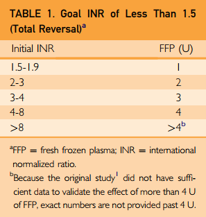
I don’t know about you, but I hate doing math in the middle of a trauma resuscitation. I need something quick and dirty. A physician from NYU Langone in NYC commented on the article, and derived a nice little table to simplify the process. He calculated the number of plasma units based on some common INR ranges, assuming that the goal was to get it down under 1.5. Here is the table:

Bottom line: This is a nice little piece of information to tuck into your pocket or phone. For patients inside the usual therapeutic values, it will take 2-3 units of plasma to reverse. For your average older human with average comorbidities, expect this to take 4-6 hours, not counting ordering, thawing, and delivery. If my definitions of “life-threatening bleeding” are met (see below), your patient may have significant adverse events during this time frame. So think very seriously about using PCC instead.

Related posts:

Reference: Fresh Frozen Plasma Dosing for Warfarin Reversal: A Practical Formula. Mayo Clin Proc 88(3):244-250, 2013.

Life Threatening Bleeding In The Anticoagulated Patient – Part 3

Over the last two posts, I’ve explored some of the current definitions of “life-threatening bleeding” and shared my own take on a simplified yet more universal definition. So how can we put this into practice?

In the trauma world, we typically need to use this definition when dealing with patients who are taking anticoagulants. When a patient on this class of drug arrives at your center after trauma, they must be evaluated promptly. This is frequently in the form of a trauma activation, which provides rapid access to labs and imaging. If the patient does not meet activation criteria, some type of expedited response (limited activation or rapid evaluation by emergency physician) is required. The most important decision that must be made is, “does this patient need to have their anticoagulant reversed?”

This decision depends on the answers to the two criteria I laid out in the last post. Is either of these present?

  1. (Physiologic) Bleeding that causes hemodynamic compromise (hemorrhagic shock) or changes in vital signs indicating progression toward it (increasing pulse rate, decreasing blood pressure).
  2. (Anatomic) Bleeding into a body region or tissues that has a high likelihood of causing death, disability, or the need for operative intervention.

The first one is easy. Actual or developing hypovolemic shock should be obvious to any clinician managing the patient.

The second one is not necessarily as apparent. Although one may think that any intracranial blood may be life-threatening, sometimes it is not. What about a little subarachnoid hemorrhage? Or a tiny subdural in an area that typically does not progress?

So how to we determine if definition 2 is met? Phone a friend. Call an expert. There are so many potential areas for this type of bleeding to occur, a single emergency physician or other clinician may not be able to accurately make this judgment. So call your friendly, neighborhood neurosurgeon (head), or surgeon (abdomen, soft tissues), GI specialist (UGI bleed), or obstetrician (baby stuff). If they agree that it is life-threatening, the reverse the anticoagulant.

This level of oversight is important, because the reversal agents are not totally benign, or cheap. They have known complications, and one rare but important one is death. So make sure that their use is justified.

Final tips: Once you have determined that reversal is required, use the fastest agent(s) available. For warfarin, this means prothrombin complex concentrate (PCC) and not plasma. Typically, plasma reversal requires at least 4 units, and this takes hours. PCC takes 30 minutes or less. 

Document your judgment well, and your conversations with specialists who are helping you with definition #2. This is critical, because there have to be checks and balances for use of your rapid reversal protocols. There must be a post hoc analysis of each and every reversal, just like there should be for use of your massive transfusion protocol. A group of knowledgeable clinicians must review the clinical information that was available at the time of presentation, and render their agreement or disagreement to provide a good feedback loop and ensure proper usage of these products.


What Is: Life Threatening Bleeding In The Anticoagulated Patient – Part 2

In my last post, I showed you some of the current definitions of “life-threatening” bleeding used in patients who are anticoagulated, as well as some for lesser degrees of bleeding. Confusing, right? How are we to ever know what is truly life-threatening so we can justify intervention/reversal with potent and/or expensive drugs and blood products?

It’s time for a more universal definition. The most important thing to recognize up front is that it is absolutely impossible to provide a comprehensive set of definitions that take every possible scenario into account. The list would be confusingly long. We really only need a few rules that are a bit more generalized. But then, how do we keep providers from just doing what they want, and coming up with some subjective or anecdotal justification? Bear with me for a day or two.

Today, I’ll lay out my two “universal definitions” of significant bleeding in an anticoagulated patient. Note that I didn’t say “life-threatening” bleeding. There is also such a thing as “limb-threatening” or “tissue-threatening” bleeding, and yet other ways to be harmed from uncontrolled bleeding due to anticoagulants.

If you look through all the various criteria that I included in the last post, you can see that they generally fall into two categories: physiologic and anatomic. The key is to reach a balance of being specific enough without being overly so. This still allows for some degree of clinician judgment. But as you will see in my next post, there also has to be some type of scrutiny and review of that judgment.

So here is what I propose. Bleeding is considered significant and/or life-threatening if either of these are true:

  1. (Physiologic) Bleeding that causes hemodynamic compromise (hemorrhagic shock) or changes in vital signs indicating progression toward it (increasing pulse rate, decreasing blood pressure).

  2. (Anatomic) Bleeding into a body region or tissues that has a high likelihood of causing death, disability, or the need for operative intervention.

In my next post, I’ll show you how to operationalize these definitions into a workable process for making decisions about reversing anticoagulation.