Best Of AAST 2022 #9: Management Of Low Grade Blunt Thoracic Aortic Injury

It has been interesting to watch the evolution of the treatment of blunt thoracic aortic injury (BTAI). In my training, patients with an abnormal mediastinal contour on chest x-ray were whisked away to interventional radiology for a thoracic aortic angiogram. Yes, there was no such thing as a CT scan for anything but the head! Here’s a sample chest x-ray:
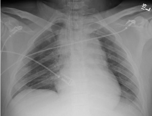
I wrote a post 12 years ago describing the various findings of a blunt aortic injury, many of which are present above. You can the post by clicking here.

In the 1-2% where the angio was positive (yes, you read that right; we did a lot of negative angiograms) the patient was then whisked off to the OR for an open aortic graft via thoracotomy. The advent of CT scans of the torso and improving resolutions allowed us to be more selective with angiography. And finally as CT angiography matured, aortic angiography became a thing of the past.

Then came TEVAR about 20 years ago. There were some growing pains as we refined the technology, but now this endovascular procedure is the standard of care for most aortic injuries. While there was also a place for nonoperative management, it was really just maintaining a reasonably low blood pressure to protect the vessel while getting the patient into good enough condition to tolerate anoperation.

Now we are beginning to slice and dice treatment based on grade. Many centers have recognized that an intimal injury (Grade I) is only a very minor disruption to the inside of the vessel and does not make it more susceptible to rupture. Grade II management has been less clear. Here is a diagram of the various grades.
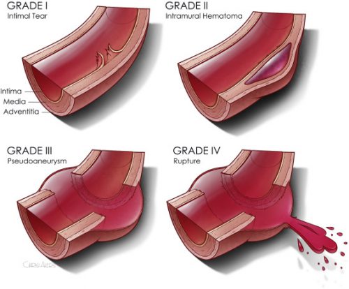
It makes sense that an invasive procedure may be less helpful for injuries that do not disrupt the layer of the vessel that provides its strength. But remember, common sense isn’t always the truth.

The group at Dell Seton Hall reviewed all patients with low grade BTAI (Grades I and II) in the Aortic Trauma Foundation Registry for a six year period. Their hypothesis was that these injuries could be successfully treated with medical management alone. They reviewed the data for mortality, complications, vent days, and lengths of stay.

Here are the factoids:

  • A total of 880 patients were enrolled and 274 had low grade injuries; 5 were then excluded when their lesion progressed and they underwent TEVAR
  • Of the 269 remaining patients, 81% were treated with medical management (81% Grade I, 19% Grade II) and the remainder with TEVAR (20% Grade I, 80% Grade II)
  • Rates of thoracotomy, craniectomy, and sternotomy were the same in both groups, but TEVAR patients were more likely to have a laparotomy (31% vs 15%)
  • Mortality was significantly higher in the TEVAR group (18% vs 8%) but the mortality from the aorta was not quite significant (4% vs 0.5%)
  • Complications (DVT and ARDS) were also significantly higher in the TEVAR group
  • Vent days and lengths of stay were equivalent

The authors concluded that medical management alone is safe and appropriate, with a lower mortality and decreased complications compared to routine TEVAR.

Bottom line: Hmm, color me skeptical. Remember, this is a registry study, so information tends to be limited outside the usual demographics and data points that are very pertinent to the purpose of the registry. The most important concept is that the patients in each group must be identical in every way except for the intervention.

We can try to make them as identical as possible by matching pairs or subgroups of patients. But the biggest problem is that the number of patient characteristics that might be important to match may not be available for analysis in the database.

When the patients were initially treated at contributing centers, there were no specific rules that the individual surgeons had to follow to decide between medical management and TEVAR. They could pick and choose based on their own experience. Could the surgeon have recognized some patients as higher risk and opted for TEVAR to make sure the aorta would not become an issue in conjunction with their other injuries? And unfortunately, perhaps that higher risk issue is what ultimately killed them and not the aorta. It’s basically a form of unhealthy user bias.

This abstract is an interesting tidbit that should push us to question whether medical management is better, at least in some subsets of low grade aortic injury patients. Then someone can perform a more robust study to confirm or refute its safety. Unfortunately, this may never happen due to the low incidence of this injury. It took six years to accumulate only 269 eligible patients in this registry!

Here are my questions for the authors and presenter:

  • What data points are actually in the registry? Specifically, is there fine detail about the other injuries the patient had? Could these have contributed to TEVAR mortality, or the selection of the patient for TEVAR to try to reduce the perceived mortality risk?
  • When you stated that there was “no difference in demographics or mechanism of injury” what were these, exactly?
  • What did patients actually die from in the Grade I and Grade II groups? Be more specific than “aortic-related” or not.
  • Do you have any worries about the five patients whose lesions progressed? When should patients be re-scanned to identify those who might benefit more from TEVAR?

This is a thought provoking abstract, and I am very interesting in hearing what the next steps should be.

Reference: MEDICAL MANAGEMENT IS THE TREATMENT OF CHOICE FOR LOW GRADE BLUNT THORACIC AORTIC INJURIES. Plenary paper #45, AAST 2022.

Best of AAST 2022 #8: Financial Vulnerability Of Trauma Centers

Here’s another abstract on trauma center / system finance. Trauma centers are part of the safety net in the healthcare systems of many countries. The way they are funded varies tremendously. In the US, health insurance pays most of the bill for patient care. Unfortunately, not all patients are covered, so there is financial risk to the center based on how many underpaying patients present for care.

The group at Scripps Mercy in San Diego performed a financial health analysis of all ACS-verified trauma centers in the US. They applied a Financial Vulnerability Score metric (FVS), although I could not locate anything on this via an internet search. They analyzed the RAND Hospital Financial Database, which is based on information obtained from the CMS Healthcare Cost Report Information System. Using this data they calculated the FVS for each center. They sub-grouped the hospitals into high, medium, and low vulnerability and compared them.

Here are the factoids:

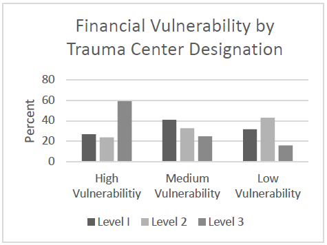
  • A total of 617 trauma centers were identified and analyzed: 194 Level I, 278 Level II, and 145 Level III
  • Level III trauma centers made up 59% of the high financial risk centers
  • The majority of Level I and II centers were in the middle or low risk categories
  • Characteristics of high risk centers were lower number of beds, negative operating margin, and less cash on hand
  • Low risk centers had greater asset to liability ratios, lower outpatient shares, and 3x less uncompensated care
  • The largest proportion of HFR hospitals were in New England and East North Central regions
  • Non-teaching centers had significantly higher financial risk than teaching hospitals (46% vs 29%)

The authors concluded that about 25% of Level I and II trauma centers are at high financial risk and that factors such as payor mix and outpatient status should be targeted to reduce this risk.

Bottom line: This is a fascinating abstract but leaves a lot to the imagination. The databases used have not been used in previous papers, and the information contained in them is proprietary. The FVS is also new and I have not been able to obtain any details.

Nevertheless, if the data and analysis are sound it may provide some new information to trauma centers and perhaps some insight on what factors to address to lessen their financial vulnerability. This is a lot of ifs. Hopefully the authors will enlighten us during the presentation so we can appreciate the real world value of the analysis.

Here are my questions and comments for the authors/presenter:

  • Please explain both the dataset used and the new FVS metric. Most readers and listeners are unfamiliar. We need to see how the data and analysis apply to trauma center financials and how the FVS has been validated.
  • How can the vulnerability factors be addressed? Payor mix is based on patient coverage and their socioeconomic status. It would seem to be difficult to manipulate by the trauma center. Outpatient vs inpatient status is also difficult to change and not fall afoul of CMS rules. What were other factors that were identified that could help centers reduce their financial vulnerability?

This could be an interesting abstract, but there was not enough room in the abstract to reveal all the details. Hopefully all will become obvious during the presentation.

Reference: FINANCIAL VULNERABILITY OF TRAUMA CENTERS: A NATIONAL ANALYSIS, Plenary paper #41, AAST 2022.

Best Of AAST 2022 #7: Funding State Trauma Systems

As of about 10 years ago, every state in the US has implemented a state trauma system. The way these systems are funded and their sources vary widely. Most receive their funds via some combination of a line item in the budget, grants, or fees levied on state residents. The total per capita funding amount can be calculated based on the total dollars received divided by the state population.

The per capita funding across the US varies widely. Some provide very little assistance while others are much more generous. I have visited trauma centers in nearly every state and have witnessed the difficulties many state trauma systems face due to funding shortfalls.

Does financial support have an impact on trauma care? A better question is, do state systems have an impact on trauma care? The literature on that last question tells us the answer is a resounding yes. We assume that having some money improves what the system can do. But that’s just an assumption. Until now.

The group at Northwestern University did a deep dive into the financial support of state trauma systems to address this question. Their goal was to measure the impact of it on something easily measurable: mortality. They selected five states to study based upon their broad geographic and demographic variability and the availability of comprehensive data for analysis.

Two states (MA, NY) allocated $0.00 per capita trauma funding, and three provided between $0.09 and $1.80 per capita.  The authors analyzed data from comprehensive patient encounter databases from these states. Their primary goal was to compare mortality differences between the two levels of support. They were also able to link ED and inpatient encounters and analyze triage decisions for appropriateness.

Here are the factoids:

  • Nearly a quarter of a million patients with ISS > 15 were analyzed over a study period of two years
  • Median age was 72 and ISS was 17 (older with significant injuries)
  • Trauma mortality was significantly decreased in the states that provided financial support compared with those that did not (odds ratio 0.75)
  • All triage levels were associated with a similar decrease in mortality (appropriate triage to a Level I or II center, undertriage admitted to a Level III or IV center, and re-triage (emergent transfer from an ED to a Level I or II center))
  • The best improvement in mortality was seen in re-triaged patients in funded states (odds ratio of 0.63)

The authors concluded that state system funding was associated with lower mortality in severely injured patients and that increasing funding may facilitate it.

Bottom line: Several papers have been written showing the benefit of having a trauma system, and all of the US has bought into this concept. However, funding of these systems varies widely by state. The assumption (up to now) has been that just having a system is enough. This abstract goes a step further and suggests that actually paying for it is important, too. 

The authors performed quite of bit of data manipulation and used some sophisticated statistical tools that I am not familiar with. However, the authors are certainly qualified, and I am going to assume that the analysis was sound. 

The one important point to keep in mind is that this paper is one of the first of its kind but can only show association, not causation. Nevertheless, it is an important contribution to the trauma system literature. It will definitely prompt additional research which will hopefully corroborate the results. This line of work should give state systems the ammunition to approach their legislatures to open the pocketbook and improve the outcomes of their residents. Mortality reductions of 25-37% cannot be ignored!

Here are my questions and comments for the authors/presenters:

  1. Please explain the data sources you used and the analysis (briefly). These are very comprehensive databases and appear to contain a wealth of data. Tell us why you chose them, how you linked them, and explain your analyses.
  2. Why limit your analyses to these five states? Could you have found some with even higher levels of support that would drive your point home even better?
  3. Be sure to provide more detail on your analysis of triage status. There are some important lessons to be learned here regarding state triage criteria vs mortality.
  4. Are you planning any additional similar studies? This is important work that will help state systems obtain a more appropriate level of support.

This is an intriguing abstract that should prompt additional research regarding state trauma system funding. 

Reference: THE IMPACT OF STATE TRAUMA FUNDING ON TRIAGE AND MORTALITY OF TRAUMA PATIENTS. Plenary paper #38, AAST 2022.

Best Of AAST 2022 #6: The “Missed” Splenic Pseudoaneurysm

Like so many things in trauma, there are two camps when it comes to repeat CT scan after solid organ injury: the believers vs the non-believers. In my experience, a minority of US trauma centers incorporate this repeat CT study in their practice guidelines. 

Yet the question keeps coming up in the literature. Earlier this year, I reviewed a paper from the University of Cincinnati from a group of believers. I was not very kind, and you can read the review here. The biggest problem with most believer papers is that they cite very old literature that overstates the incidence of delayed hemorrhage. They then use this to justify an extra CT scan to find more of these “dangerous” pseudoaneurysms. Unfortunately, those old papers are just not very good and many overstate the problem.

So let’s look at this year’s abstract from the LAC+USC group. They open by stating that the natural history is unclear but that “risk for spontaneous rupture and exsanguination exist.” The authors sought to further define the utility of using a delayed CT angiogram (dCTA) in diagnosing and triggering intervention after high-grade blunt solid organ injury.

They performed a retrospective study of all patients arriving at their Level I center over a nearly five year period with a Grade 3 or higher injury to liver, spleen, or kidney. They excluded the young, patients transferred in, early deaths, and patients who underwent immediate operation on their spleen or kidney. The primary outcome was intervention triggered by the dCTA.

Here are the factoids:

  • A total of 349 patients with 395 high grade solid organ injuries were analyzed (42% liver, 30% spleen, 28% kidney)
  • Median injury grade for each organ was 3
  • Initial management was “typically” nonoperative or angioembolization (liver 83%, spleen 95%, kidney 89%)
  • Delayed CT angiogram was typically performed on day 4 and identified a lesion in 16 spleen, 10 liver, and 6 renal injuries
  • The dCTA prompted an intervention in 12 spleen, 8 liver, and 5 kidney injuries

The authors conclude that delayed CTA identified a significant number of vascular lesions requiring endovascular or surgical intervention. They recommend further examination and consideration of universal screening to avoid missing these pesky pseudoaneurysms.

Bottom line: Once again, we have a paper that conflates finding a pseudoaneurysm with the need to get rid of it. Granted, I was always taught that pseudoaneurysms (in adults) found on initial CT required an intervention. In the old days of “delayed splenic rupture” a pseudoaneurysm was the likely culprit. 

But the majority of centers do not go looking for pseudoaneurysms days later. And there are precious few patients coming back with delayed hemorrhage after discharge. So what gives?

Could it be that there is a difference between a “fresh” pseudoaneurysm and a “delayed” one? Perhaps the fresh ones portend a real risk of bleeding, but delayed ones are just a normal part of the healing process and rarely bleed? We just don’t know for sure.

This paper shows that if you look for a delayed pseudoaneurysm you will find them. And at this center, if you find them you will be compelled to angioembolize or even operate on them. Yet we really don’t know if that is necessary. It certainly adds to length of stay and hospital charges.

My take is that we desperately need a broad tally of patients discharged with a liver or spleen injury who return within a few weeks for bleeding complications. I would exclude kidneys because they act so differently. And I would not look at all returns because most liver injury readmissions are for bile problems. Just focus on readmissions for bleeding. Once we see what the real incidence is, we can decide whether these pseudoaneurysms are a problem significant enough to pursue with delayed scans, etc.

Here are my questions for the authors and presenter:

  1. What is your assessment of the incidence of delayed rupture and exsanguination? Have you read through the old papers in detail to assure yourselves that they are actually correct?
  2. Do you hold patients in the hospital for their delayed CT angiogram? The studies were typically performed on days 3-7. Do you really keep your solid organ injured patients in the hospital that long? At our center, a grade 3 injury could be discharged home in two days!
  3. How do you decide to take a patient to interventional radiology or the OR after the delayed CT? Is it an unwritten rule? It seemed like most, but not all, had some type of intervention. A (very) few had the lesion but nothing was done. Please explain the difference.

This is an interesting paper just because of the intuitive leap it makes from pseudoaneurysm to intervention. I’m anticipating your presentation so I can hear all the details.

Reference: PSEUDOANEURYSMS AFTER HIGH GRADE BLUNT SOLID ORGAN INJURY AND THE UTILITY OF DELAYED CT ANGIOGRAPHY. Plenary paper #34, AAST 2022.

Best Of AAST 2022 #5: Traumatic Subarachnoid Hemorrhage

Traditionally, just about any type of intracranial hemorrhage (ICH) prompts a thorough neurological investigation and frequently involves a neurosurgery consultation. Subarachnoid hemorrhage (SAH) is one type of ICH that has historically demonstrated very few adverse consequences. Yet the custom of performing a time-consuming and expensive evaluation has persisted.

Over the past few years, the literature about the lack of utility of this practice has been growing. Many trauma centers (including my own at Regions Hospital) have simplified the evaluation of traumatic SAH. We have actually come to a point that we don’t even admit most of these patients. And if we are not going to admit them, why should we even accept them in transfer from other hospitals?

The trauma group at Lehigh Valley in Pennsylvania performed a five-year review of their own hospital experience (Level I trauma center) from 2015-2019. They wanted to answer the question about the necessity of transferring SAH patients to a higher-level trauma center for evaluation. They limited the study to patients with an isolated SAH. These patients were evaluated for factors that would indicate the potential for a worsening repeat CT scan.

Here are the factoids:

  • The study patients were older (mean 73 years) with a slight preponderance of females
  • ISS was low (mean 5) as would be expected for an isolated injury
  • Slightly more than half (57%) were transferred from other hospitals
  • A total of 350 patients were reviewed, and 97% had a neurosurgery consult
  • Only one patient required neurosurgical intervention, and four died in hospital
  • A total of 311 patients had a repeat CT scan and 16 showed a worse result
  • Higher AIS head was associated with a worse result

The authors concluded that it is safe to manage traumatic SAH without transfer to a higher level trauma center.  They also suggest that neurosurgical consultation is not needed and that repeat imaging may not be very useful.

Bottom line: This abstract conforms to my own opinion, so I have to take great care not to succumb to my confirmation bias. The authors found that nearly no one needed neurosurgical intervention and concluded that a neurosurgical consult is probably not needed. This is all based on a low incidence of progression and few adverse events. I agree totally.

But I also think that referring hospitals will find these few adverse events and progressions troubling. Remember, 16 patients had a worse scan,  four died during their hospital stay, and one needed an operation. Thus, it will be very important for the authors to explain the details of these patients to assuage any fears that they might deteriorate in the outside hospital ED. 

My own experience indicates that “worsening CT” is typically just a slightly larger SAH and not the sudden development of subdural or epidural blood. This is more likely to happen in patients on thinners and they should be excluded from this pool anyway. They have very specific evaluation needs.

Based on recent published literature and our own clinical experience, the Regions trauma program discharges any patient with traumatic SAH who is not on thinners and has a GCS 15. They are given instructions to follow up with their primary care provider, and to schedule a TBI clinic appointment if they feel they have any persistent post-concussive symptoms. And because of this practice, the ED will not accept transfer of patients with this isolated injury.

You can download a copy of the protocol here.

Here are my questions for the authors and presenter:

  1. Please provide details on the patients with CT progression, operation, and death. We need to know if these were serious CT changes, or just incidental findings and complications from other causes.
  2. How successful have you been at resisting transfers in? Most referring centers are initially “not comfortable” with these patients, even though they can easily follow our simple evaluation guideline. It takes time and education to bring them into the fold.

I enjoyed this abstract and await the details to come in the presentation!