How Quickly Does Hemoglobin Drop After Acute Bleeding?

We all know that hemoglobin / hematocrit drop after blood loss. We can see it decreasing over the days after acute bleeding or a major operative procedure (think orthopedics). And we’ve been told that the hemoglobin value doesn’t drop immediately after acute blood loss.

But is it true? Or is it just dogma?

A reader sent me a request for some hard references to support this. When I read it, I knew I just had to dig into it. This is one of those topics that gets preached as dogma, and I’ve bought into it as well.

Now, I have personally observed both situations. Long ago, I had a patient with a spleen injury who was being monitored in the ICU with frequent vital signs and serial blood draws (but I don’t do that one anymore). He was doing well, then became acutely hypotensive. As he was being whisked off to the OR, his most recent hemoglobin came back at 10, which was little changed from his initial 11.5 and certainly no independent reason to worry.

But hypotension is a hard fail for nonoperative solid organ management. In the OR, anesthesia drew another Hgb at the end of the case, and the value came back 6.

Similarly, we’ve all taken care of patients who have had their pelvis fixed and watched their Hgb levels drop for days. Is this anecdotal or is it real? The doctor / nursing / EMS textbooks usually devote about one sentence to it, but there are no supporting references.

I was only able to locate a few older papers on this. The first looked at the effect of removing two units of red cells acutely. Unfortunately, the authors muddied the waters a little. They were only interested in the effect of the lost red cell mass on cardiac function, so they gave the plasma back. This kind of defeats the purpose, but it was possible to see what happened to Hgb levels over time.

Here were there findings over time for a group of 8 healthy men:

Time Hbg level
Before phlebotomy 14.4
1 week after 11.7
4 weeks after 12.6
8 weeks after 13.6
16 weeks after 13.9

So the nadir Hgb value occurred some time during the first week after the draw and took quite some time to build back up from bone marrow activity.

That’s the longer term picture for hemoglobin decrease and return to normal. What about more acutely? For this, I found a paper from a group in Beijing who was trying to measure the impact of Hgb loss from a 400cc blood donation on EEG patterns. Interesting.

But they did do pre- and post-donation hemoglobin values. They found that the average Hgb decreased from 14.0 to 13.5 g/dl during the study, which appeared to be brief. Unfortunately, this was the best I could find and it was not that helpful.

Bottom line: Your patient has lost whole blood. So, in theory, there should be no Hgb concentration difference at all. But our bodies are smart. The kidneys immediately sense the acute hypovolemia and begin retaining water. The causes ongoing hemodilution within seconds to minutes. Additionally, fluid in the interstitial space begins to move into the vascular space to replace the volume lost. And over a longer period of time, if no additional fluid is given the intracellular water will move out to the interstitium and into the vascular space.

But these things take time. There is an accelerating curve of hemodilution that takes place over hours. The slope of that curve depends on how much blood is lost. A typical 500cc blood transfusion will cause a 0.5 gm/dl drop over several minutes to an hour. We don’t have great data on the exact time to nadir, but my clinical observations support a hyperbolic curve that reaches the lowest Hgb level after about 3 days.

Unlike this curve, it levels off and slowly starts to rise after day 3-4 due to bone marrow activity.

The steepness of the curve depends on the magnitude of the blood loss. After a one unit donation, you may see a 0.5 gm/dl drop acutely, and a nadir of 1 gm/dl. In the case of the acutely bleeding patient with the spleen injury, the initial drop was 1.5 gm/dl. But two hours later it had dropped by over 5 gm/dl. 

Unfortunately, the supporting papers are weak because apparently no one was interesting in proving or disproving this. They were more interested in cardiac function or brain waves. But it does happen. 

Here’s the takeaway rule:

In a patient with acute bleeding, the initial hemoglobin drop is just the tip of the iceberg. Assume that this is only a third (or less) of how low it is going to go. If it has fallen outside of the “normal” range, call for blood. You’ll need it!

References:

  1. Effect on cardiovascular function and iron metabolism of the acute removal of 2 units of red cells. Transfusion 34(7):573-577, 1994.
  2. The Impact of a Regular Blood Donation on the Hematology
    and EEG of Healthy Young Male Blood Donors. Brain Topography 25:116-123, 2012.


What You Need To Know About: Frontal Sinus Fractures

Fracture of the frontal sinus is less common than other facial injuries, but can be more complex to deal with, both in the shorter and longer terms. These are generally high energy injuries, and facial impact in car crashes is the most common mechanism. Fists generally can’t cause the injury, but blunt objects like baseball bats can.

Here’s the normal anatomy:

sinus-fracture-treatment


Source: www.facialtraumamd.com

There are two “tables”, the anterior and the posterior. The anterior is covered with skin and a small amount of subcutaneous tissue. The posterior table is separated from the brain by the meninges.

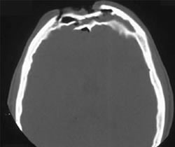
Here’s an image of an open fracture involving both tables. Note the underlying pneumocephalus.

frontal_sinus1

A third of injuries violate the anterior table, and two thirds violate both. Posterior table fractures are very rare. A third of all patients will develop a CSF leak, typically from their nose.

These fractures may be (rarely) identified on physical exam if deformity and flattening is noted over the forehead. Most of the time, these patients undergo imaging for brain injury and the fracture is found incidentally. Once identified, go back and specifically look for a CSF leak. Clear fluid in the nose is, by definition, CSF. Don’t waste time on a beta-2 transferring (see below).

If a laceration is clearly visible over the fracture, or if a CSF leak was identified, notify your maxillofacial specialist immediately. If more than a little pneumocephalus is present, let your neurosurgeon know. Otherwise, your consults can wait until the next morning.

In general, these patients frequently require surgery for the fracture, either to restore cosmetic contours or to avoid mucocele formation. However, these are seldom needed urgently unless the fracture is an open fracture with contamination or there is a significant CSF leak. If in doubt, though, consult your specialist.

Related posts:

New Technology: Using AI To Interpret Pelvic X-rays

Look out, radiologists! The computers are coming for you!

Radiologists use their extensive understanding of human anatomy and combine it with subtle findings they see on x-ray shadow pictures. In doing this, they can identify a wide variety of diseases, anomalies, and injuries. But as we have seen with vision systems and game playing (think chess), computers are getting pretty good at doing this as well.

Is it only a matter of time until computer artificial intelligence (AI) starts reading x-rays?  Look at how good they already are at interpreting EKGs. The trauma group at Stanford paired up with the Chang Gung Memorial Hospital in Taiwan to test the use of AI for interpreting images to identify a specific set of common pelvic fractures.

The Stanford group used a deep learning neural network (XCeption) to analyze source x-rays (standard A-P pelvis images) from Chang Gung. These x-rays were divided into training and testing cohorts. The authors also applied different degrees of blurring, brightness, rotation, and contrast adjustment to the training set in order to help the AI overcome these issues when interpreting novel images.

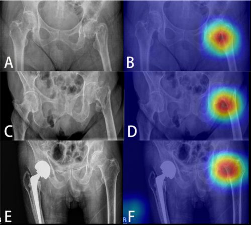
The AI interpreted the test images with a very high degree of sensitivity, specificity, accuracy, and predictive values, with all of them over 0.90. The algorithms generated a “heat map” that showed the areas that were suspicious for fracture. Here are some examples with the original x-ray on the left and the heat map on the right:

The top row shows a femoral neck fracture, the middle row an intertrochanteric fracture, and the bottom row another femoral neck fracture with a contralateral implant. All were handily identified by the AI.

AI applications are usually only as good as their training sets. In general, the bigger the better so they can gain a broader experience for more accurate interpretation. So it is possible that uncommon, subtle fractures could be missed. But remember, artificial intelligence is meant to supplement the radiologist, not replace him or her. You can all breathe more easily now.

This technology has the potential for broader use in radiographic interpretation. In my mind, the best way to use it is to first let the radiologist read the images as they usually do. Once they have done this, then turn on the heat map so they can see any additional anomalies the AI has found. They can then use this information to supplement the initial interpretation.

Expect to see more work like this in the future. I predict that, ultimately, the picture archiving and communications systems (PACS) software providers will build this into their product. As the digital images are moving from the imaging hardware to the digital storage media, the AI can intercept it and begin the augmented interpretation process. The radiologist will then be able to turn on the heat map as soon as the images arrive on their workstation.

Stay tuned! I’m sure there is more like this to come!

Reference: Practical computer vision application to detect hip fractures on pelvic X-rays: a bi-institutional study.  Trauma Surgery and Acute Care Open 6(1), http://dx.doi.org/10.1136/tsaco-2021-000705.

Do I Have To Call My Trauma Team For Incoming Transfers?

I had a great question sent in by a reader last week:

Some trauma centers receive a number of transfers  from referring hospitals. Much of the time, a portion of the workup has already been done by that hospital. If the patient meets one or more of your trauma activation criteria, do you still need to activate your team when they arrive?

And the answer is: sometimes. But probably not that often.

Think about it. The reason you should be activating your team is that you suspect the patient may have an injury that demands rapid diagnosis and treatment. The purpose of any trauma activation is speed. Rapid evaluation. Fast lab results. Quick access to CT scan or OR. If a significant amount of time has already passed (transported to an outside hospital, worked up for an hour or two, then transported to you), then it is less likely that a trauma activation will benefit the patient.

There are four classes of trauma activation criteria. I’ll touch on each one and the need to activate in a delayed fashion if present, in priority order.

  • Physiologic. If there is a significant disturbance in vital signs while in transit to you (hypotension, tachycardia, respiratory problems, coma), then you must activate. Something else is going on that needs to be corrected as soon as the patient arrives. And remember the two mandatory ACS criteria that fall into this category: respiratory compromise/need for an emergent airway, and patients receiving blood to maintain vital signs. But a patient who needed an airway who is already intubated and no longer compromised does not need to be a trauma activation.
  • Anatomic. Most simple anatomic criteria (e.g. long bone or pelvic fractures) do not need a trauma activation unless the patient is beginning to show signs of physiologic compromise. However, anatomic criteria that require rapid treatment or access to the OR (proximal amputations, mangled or pulseless extremities, spinal cord injury) should be activated.
  • Mechanism. Most of the vague mechanistic criteria (falls, pedestrian struck, vehicle intrusion) do not require trauma activation after transfer to you. But once again, if the mechanism suggests a need for further rapid diagnosis or treatment (penetrating injury to abdomen), then activate.
  • Comorbidities. This includes underlying diseases, extremes of age, and pregnancy. In general, these will not require trauma activation after they arrive.

Bottom line: In many cases, the patient transferred in from another hospital will not need to be a trauma activation, especially if they have been reasonably assessed there. The patient should be rapidly eyeballed by your emergency physicians, and if there is any doubt about their condition, activate then.

However, if little workup was done at the outside hospital (my preference), and the injuries are “fresh” (less than a few hours old), then definitely call your team. 

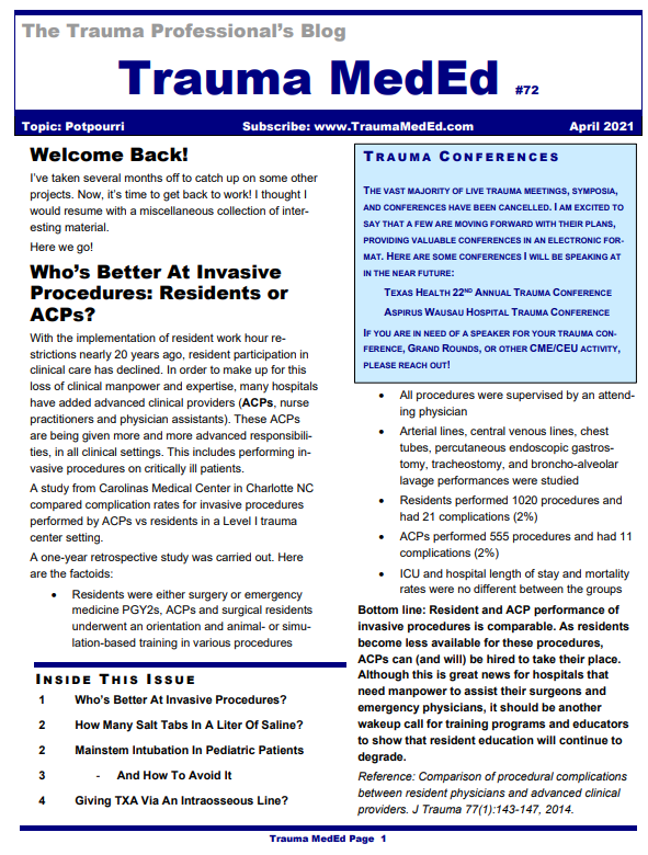
The April 2021 Trauma MedEd Newsletter Is Live! Potpourri

This issue is devoted to an uncommon yet potentially devastating problem, blunt carotid and vertebral artery injury.

In this issue, learn about:

  • Who’s Better At Invasive Procedures? Advanced care providers or residents?
  • How Many Salt Tabs In A Liter Of Saline?
  • Mainstem Intubation In Pediatric Patients
  •    And How To Avoid It!
  • Giving TXA Via An Intraosseous Line?

To download the current issue, just click here!

Or copy this link into your browser: https://bit.ly/TME202104

This newsletter was released to subscribers over a week ago. If you would like to be the first to get your hands on future newsletters, just click here to subscribe!