This xray is a classic for a specific trauma surgical injury. Give it your best shot! Tweet of leave comments with your impressions.
This image is especially appropriate for surgical residents / registrars.
Answer in the next post!
This xray is a classic for a specific trauma surgical injury. Give it your best shot! Tweet of leave comments with your impressions.
This image is especially appropriate for surgical residents / registrars.
Answer in the next post!
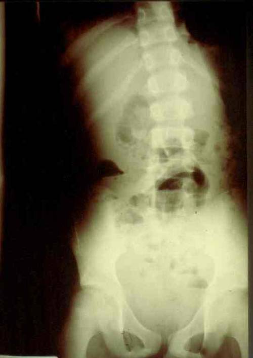
Blunt injury to hollow organs is rare in adults, but a little more common in children. This is due to their smaller muscle mass and the lack of protection by their more flexible skeleton. Duodenal injury is very rare, and most trauma professionals don’t see any during their career. As with many pediatric injuries, there has been a move toward nonoperative management in selected cases, and duodenal injury is no exception.
What we really need to know is, which child needs prompt operative treatment, and which ones can be treated without it? Children’s Hospital of Boston did a multicenter study of pediatric patients who underwent operation for their injury to try to tease out some answers about who needs surgery and what the consequences were.
A total of 16 children’s hospitals participated in this 4 ½ year study. Only 54 children had a duodenal injury, proven either by operation or autopsy. Some key points identified were:
Although laparoscopic exploration was attempted in about 12% of patients, it was universally converted to an open procedure when the injury was confirmed. TPN was used commonly in the postop period. Postop ileus was very common, but serious complications were rare (wound infection <10%, abscess 3%, fistula 4%). There were 2 deaths: one child presented in extremis, the other deteriorated one day after delayed recognition of the injury.
Bottom line: Be alert for this rare injury in children. Marks on the abdomen, particularly the epigastrium, should raise suspicion of a duodenal injury. The best imaging technique is the abdominal CT scan. Contrast is generally not helpful and not tolerated well by children. Duodenal hematoma can be managed nonoperatively. But any evidence of perforation (free fluid, air bubbles in the retroperitoneum, duodenal wall thickening, elevated serum amylase) should send the child to the OR. And laparotomy, not laparoscopy, is the way to go.
Reference: Operative blunt duodenal injury in children: a multi-institutional review. J Ped Surg 47(10):1833-1836, 2012.
Let’s look at an uncommon scenario that crops up from time to time. Most seasoned trauma professionals have seen this one a time or two:
An elderly male is driving on a sunny afternoon, and crashes his car into a highway divider at 25 miles per hour. EMS responds and notes that he has a few facial lacerations, is awake but confused. They note some possible facial asymmetry and perhaps a bit of upper extremity weakness. No medical history is available. Witnesses state that he was driving erratically before he crashed. Medics call the receiving trauma center in advance to advise them that they have a stroke code.
Is this a reasonable request? Stroke centers pride themselves on the speed of their stroke teams in assessing, scanning, and when appropriate, administering thrombolytics to resolve the problem. But if there are suspicions of stroke in a trauma patient, which diagnosis wins? Trauma team or stroke team?
Lets analyze this a bit further, starting with diagnosis. Remember the first law of trauma:
Any anomaly in your trauma patient is due to trauma, no matter how unlikely it may seem.
Could the symptoms that the paramedics are observing be due to the car crash? Absolutely! The patient could have a subdural or epidural hematoma that is compressing a cranial nerve. There might be a central cord injury causing the arm weakness. His TBI might be the source of his confusion. The facial asymmetry could be due to a pre-existing Bell’s palsy, or he could have had a stroke years ago from which he has only partially recovered.
If the stroke team is called for the patient, they will focus on the neuro exam and the brain. They will not think about trauma. They will follow the patient to CT scan looking for the thing that they do best with. If they don’t see it, the patient will return to the ED for (hopefully) a full trauma workup. If there are occult injuries in the abdomen, then the patient may have been bleeding for an hour by then. This elderly patient will then be way behind the eight ball.
And let me pose the worst case scenario. The patient is taken to CT by the stroke team, and lo and behold he has a thrombotic stroke! This patient had a stroke, which caused him to lose control of his car and explains most of his findings. Again, the stroke team will do what they are trained to do and give a thrombolytic. They are still not thinking about trauma. Within minutes the patient becomes hypotensive and his abdomen appears a bit more distended. He is rushed back to the ED (remember, no CT in hypotensive patients even if you are in the scanner) and a FAST exam is very positive for free fluid throughout the abdomen. Imagine the look you will get from the surgeon as they run to the OR to perform a splenectomy on this fully anticoagulated patient!
Bottom line: If you have a patient who is trauma vs stroke, trauma always wins! Remember the first law and try to find traumatic reasons for all signs and symptoms. Perform your standard trauma workup and incorporate the appropriate head scans into your evaluation. Then and only then should the stroke team be called.