Secondary Overtriage: What Is It, And Why Is It Bad?

Simply put, secondary overtriage (SO) is the unnecessary transfer of a patient to another hospital. How can you, as the referring trauma professional, know that it is unnecessary? Almost by definition, you can’t, unless you have some kind of precognition. If you knew it wasn’t necessary, you wouldn’t do it in the first place, right?

But using the retrospectoscope, it’s much easier. The classic definition describes a patient who is discharged from the hospital shortly after arrival there. What is “shortly?” Typically, it occurs within 48 hours in a patient with low injury severity (ISS < 16) and without operative intervention. Definitions may vary slightly.

And why is it bad?

Several states with rural trauma systems have scrutinized this issue. The first study is from West Virginia, where six years of state registry data were analyzed. Over 19,000 adults were discharged home from a non-Level I center within 48 hours after an injury. Of those, about 1,900 (10%) had been transferred to a “higher level of care” and discharged from that center (secondary overtriage, could be any higher-level trauma center).

The factoids:

  • Patients with ISS > 15 and requiring blood transfusion were more likely to be SO. (I would argue that this is appropriate triage in most cases!)
  • Neurosurgical, spine and facial injuries were also associated with SO. (This one is a little more interesting, see below).
  • SO was more likely for transfers during the night shift, when resources are often more scarce

The problem is that this study is descriptive only. It doesn’t really help us figure out which patients could/should be kept based on any of the variables they collected.

The next study is from Dartmouth in New Hampshire and examines transfers into that single Level I center from 72 other hospitals. Registry data were examined over 5 years, identifying transfer patients with ISS < 15 who were discharged within 48 hours without an operation.

Yet more factoids:

  • 62% of the nearly 8,000 patients received by this center were transfers
  • Overall SO rate was 26%
  • A quarter of adult patients and one half of pediatric patients were considered SO, and about 15% of them were actually discharged from the ED (!)
  • Head and neck, and soft tissue injuries were most common among SO patients

The real bottom line: Here are my thoughts on what you can do to try to decrease the number of your patients with SO and optimize the transfer process:

  • Work with your upstream trauma center to determine how much imaging you really need to perform
  • Develop a reliable method of getting those images to them
  • Ask them to help you develop practice guidelines and educate your hospital/ED staff to help manage common diagnoses that often result in SO from your center
  • If you are located in a rural area, inquire about RTTD courses you might attend

References:

  • Secondary overtriage in a statewide rural trauma system. J Surg Research 198:462-467, 2015.
  • Secondary overtriage: the burden of unnecessary interfacility transfers in a rural trauma system. JAMA Surg 48(8):763-768, 2013.

Radiographic Image Sharing Systems

There are generally three ways to share radiographic images with your upstream trauma center:

  • Hard copy. These days, that usually means a CD. Nearly all PACS systems (picture archiving and communications systems) can write CDs that can accompany your patient. Advantage: super cheap. Possible downsides: the CD may be corrupted and not openable, the software on the disk cannot be installed or will not run at the receiving hospital, and finally it can just be forgotten in the rush to get the patient out of the ED.
  • PACS system connections. These are software links that enable one hospital’s PACS software to communicate with another’s. They must be established in advance, and generally require some expertise from the hospitals’ IT departments. Images can be pushed from one system to another. Advantages: once set up, it is very inexpensive to maintain, and images can be viewed prior to patient arrival at the receiving hospital. Possible downside: Al-though the interchange format is standardized, every once in a while the systems just can’t communicate.
  • Web-based image sharing system. This consists of a web server-based software application available via the internet that allows subscribing hospitals to sign on and share images. Referring hospitals can upload images from their PACS systems for free, and the receiving hospital can view the images and/or download into their own system. Advantage: these products are simple to set up, and easy to use after just a little training. Compatibility is very high, and the services are continually working to ensure it. Downside: expensive. Depending on specifics, the annual subscription may be up to $100K per year, and is generally footed by the receiving trauma center.

Is a web-based solution worth it? MetroHealth in Cleveland looked at this over five years ago, and published their results in 2015. They looked at their experience pre- and post-implementation and found the following:

  • Three years of transfer data prior to the web system implementation was compared to one year of experience after
  • CT imaging decreased at both referring and receiving hospitals across the study period
  • Repeat scan rate decreased from 38% to 28%. Repeat head scans were the major driver at 21%.
  • Cost of reimaging dropped from about $1000 per patient to $600

Bottom line: As a referring hospital, it is your responsibility to ensure that the (hopefully) few images you obtain make it to the upstream trauma center. Although hard copy (CD) is the cheapest, it is also the least reliable. Work with your radiology and IT departments to determine which electronic solution is best for you. Some states and regional trauma systems help subsidize or provide a web-based solution for their member hospitals.

Reference: Implementation of an image sharing system significantly reduced repeat computed tomographic imaging in a regional trauma system. J Trauma 80(1):51-56, 2016.

Impact Of Patient Imaging Prior To Transfer Out

The reality is that 90% of injuries are minor and can be treated at any hospital. A minority of patients actually have issues that require transfer to a higher-level trauma center. Physical examination can certainly help determine who some of those patients are. Think obvious open fracture or severe brain injury at a hospital without key specialists to care for them.

But not all injuries are that obvious. Imaging techniques are the next step to identifying injuries that would require transfer. The question is, how much imaging is appropriate?

A few hospitals are selective about it. But many proceed with a comprehensive battery of scans and x-rays. Some believe that their receiving trauma center expects it. And a few may be doing it for the money, unfortunately. So who is right?

There are three issues at play: time, accuracy, and radiation exposure. Let’s pick them apart.

Time. It takes time to get radiographic studies. Depending on the number obtained, it can take up to 90 minutes. A study looking at transfers from rural hospitals to a regional trauma center in Wisconsin found that the median time to transfer significantly in-creased from 67 to 140 minutes with the addition of even a single CT scan.

This issue appears to be even more of a problem in children. A group at Cincinnati Children’s Hospital studied the characteristics of children who experienced prolonged transfer times to a Level I pediatric trauma center. They reviewed 5 years of registry data, looking at time of injury to time of arrival at their center. The State of Ohio has a goal of a maximum 2-hour transfer time.

And here are the factoids:

  • 748 patients were included in the study, and the demographics were predictable (65%male, 97% blunt)
  • 25% were more severely injured (ISS > 15)
  • The majority of the patients (82%) arrived well after the 2-hour goal (7 hrs!!)
  • 79% of patients with high ISS and 47% of those with severe TBI arrived late (!!)
  • Transfer tardiness did not correlate with distance, and was only slightly improved when a helicopter was used
  • Significantly more CT scans were obtained in the late transfer group (49% vs 23%), and appeared to have no correlation with GCS or vital signs. There was, however, a significant correlation with private insurance.
  • Half of the children with scans arrived without results or had suboptimal imaging, resulting in repeat scans in about one third.

Accuracy and radiation exposure. These two factors are inextricably linked because inaccuracy begets additional imaging. As noted in the previous study, radiology results are frequently lacking, or the studies are not done well, as determined by the receiving center. This means that inaccurate results, or no results at all, are available after transfer. How much of a problem is this?

The Level I center at UC Davis looked at all incoming trauma transfers that had any CT imaging done prior. Of 370 patients, one quarter needed repeat scans. Most common were head scans (47%) and cervical spine (20%). The most common reasons for repetition were referring hospital scan not available (42%) (not sent, couldn’t open) and insufficient quality (20%). This resulted in significant additional radiation exposure, with 4% of patients receiving more than 10mSv!

Bottom line: Imaging prior to transfer definitely increases time to transfer and frequently results in repeat imaging and more radiation exposure. So why does it happen? Sometimes, it’s the mistaken belief that it will save time after transfer. Not the case. Or there is time left before the transport ambulance or helicopter arrives, so why not use it? Not a good reason, and it may delay the transfer team if they arrive early. Or the receiving trauma center “expects it.” Not if they’ve looked at any of these papers!

The best approach is to order only images that will guide your therapy. A chest x-ray on arrival or after intubation. A pelvic x-ray to determine if a binder should be applied. A CT of the abdomen to see if there are any injuries that can’t be taken care of at your hospital. As a general rule, once you have found an injury your hospital can’t treat, or have made the decision to transfer for any other reason, no additional imaging is indicated! 

References:

  • Consequences of increased use of computed tomography imaging for trauma patients in rural referring hospitals prior to transfer to a regional trauma centre. Injury 45:835-839, 2014.
  • Unnecessary imaging, not hospital distance, or transportation mode impacts delays in the transfer of injured children. Pediatric Emerg Care 26(7):481-486, 2010.
  • Rate and Reasons for Repeat CT Scanning in Transferred Trauma Patients. Am Surg 83(5):465-569, 2017.

Rural Trauma Team Development Course Impact On Trauma Transfers

The Rural Trauma Team Development Course (RTTDC) is yet another quality program developed by the American College of Surgeons (ACS). It is designed for all trauma professionals in rural areas including doctors, nurses, advanced practice providers, prehospital providers, and administrative support. The course is presented over the course of one day and covers a number of topics including:

  • Organizing a rural trauma team
  • Preparing rural hospitals to manage trauma patients
  • Identifying local resources and limitations
  • Resuscitation of trauma patients
  • Initiating early transfer
  • Developing a performance improvement process
  • Building relationships between rural hospitals and regional or state trauma systems

The trauma group at Vanderbilt compared a group of six non-trauma hospital in rural Tennessee who had participated in the RTTDC with six other rural hospitals matched for size, volume, and distance from the Level I center.

Here are the factoids:

  • A total of 130 RTTDC patients were compared with 123 from hospitals that had not participated
  • Overall demographics and number of imaging studies were the same
  • The call to transfer occurred 41 minutes sooner in the RTTDC hospitals
  • Length of stay in the referring ED was 61 minutes shorter in the RTTDC hospitals
  • Number of images obtained pre-transfer and mortality were unchanged

Bottom line: The numbers were small and the review was retrospective, but the results are nonetheless impressive. Granted, there was no decrease in mortality, but this is a relatively crude indicator, especially when small numbers are involved. But time to phone call and time spent in the referring ED were significantly shorter. Does anyone think that longer times to transfer are somehow good for patients?

Rural hospitals should consider attending RTTDC in order to improve the care of patients from their communities.

Reference: Rural trauma team development course de-creases time to transfer for trauma patients. J Trauma 81(4):632-637, 2016.

Are You Still Using MRI To Clear The Cervical Spine?

There is a fairly robust  amount of data that shows that, properly performed, the cervical spine can be cleared using a high quality CT read by a highly skilled radiologist. This is true even for obtunded patients. Pooled data suggest that the miss rate in this group is only 0.017%. And MRI is not perfect either, missing significant ligamentous injury in a small number of patients.

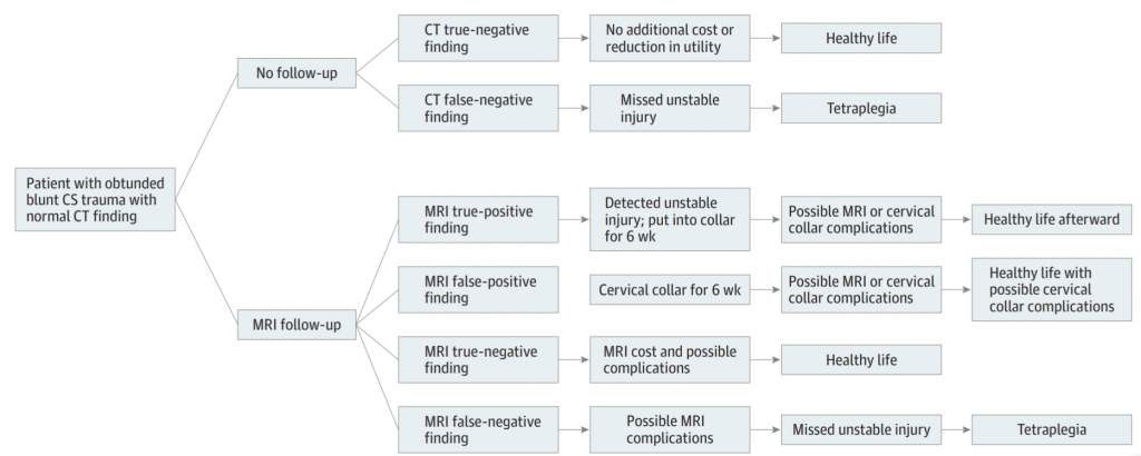
But it seems that some trauma professionals are still using MRI in some cases despite this data. The latest study on MRI focuses on the cost-effectiveness of the technique. The authors selected patients with GCS < 13 to be their obtunded group, which is probably a bit high. Nevertheless, they used a fairly sophisticated (meaning hard to understand) modeling-based decision analysis using a computerized simulation. This allowed them to compare different clearance strategies without performing large randomized clinical trials.

The authors considered MRI vs no MRI, false results, collar use and complications, MRI use with cost and complications, and the worst-case scenario of tetraplegia. Here is a flow chart of the scenarios considered. (Courtesy JAMA Surgery)

Here are the factoids:

  • The mean cost for followup vs no followup was $14K vs $1K, with no increase in quality adjusted life years (QALY)
  • No followup was the better strategy when the negative predictive value of CT was high (>98%), when the risk of an unstable injury treated with a collar turning into a permanent deficit was >25%, or if the chance of a missed injury becoming a permanent deficit was >58%
  • No followup MRI was the better strategy in all 10,000 iterations of the simulation

Bottom line: Yes, this is a fairly heavy computer simulation. But the reality is that we will never be able to design a large enough study to critically evaluate this issue and have it pass any IRB review. So it’s probably as good as it will ever get. It’s time to stop wasting money and putting obtunded patients in harm’s way by locking them into a relatively inaccessible MRI scanner for 30 minutes just to confirm the CT. Or keeping a collar until until the skin breaks down.

Here is a copy of the practice guideline we use for clearing all cervical spines, obtunded or not. Yes, there is some weirdness with soft collars, which mainly serve as a reminder to re-examine the patient at some point. But note the scan technique and requirement that it be read by a neuroradiologist for final clearance.

Related link:

Reference: Cost-effectiveness of Magnetic Resonance Imaging in Cervical
Clearance of Obtunded Blunt Trauma After a Normal
Computed Tomographic Finding