Practical Tips: Transferring The Mangled Extremity

Managing the mangled extremity is both challenging and intense. There is always pressure to do all we can to save that threatened limb. But as you know, different levels of trauma centers have different capabilities and specialists that are needed to fully manage these injuries.

Level I centers have a comprehensive set of specialists to deal with the managed extremity, including trauma surgeons, vascular surgeons, orthopedic surgeons comfortable with complex injury, plastic surgeons, and interventional radiologists. The expectation is that a mangled extremity can be completely managed at such a center.

Level III centers have much more limited resources, and may only have a trauma surgeon to perform the initial evaluation. Definitive management can only occur after transfer to a Level I center.

Level II centers often find themselves in a kind of limbo. They have most of the specialties required, but those specialists may have varying comfort levels regarding addressing complex injuries. Some Level II centers may be able to keep these patients, but many will find that they need to transfer to their upstream Level I partner.

What do transferring trauma centers need to do before actually moving the patient? Here are some practical tips.

  • Evaluate quickly. The bottom line is to try to preserve function, so time is of the essence. Do a thorough evaluation of the anatomy, as well as vascular and neurologic status. These are the major determinants of salvageability.
  • Don’t ignore the rest of the patient. Make sure that injuries more critical than the extremity are identified and addressed. See the “Dang Factor!” below.
  • Make a decision. Now. Decide whether you need to transfer the patient based on your knowledge of your consultants’ skill levels and comfort.
  • Once you decide you will transfer, do no further imaging. It’s not going to change anything you do, and may not be very helpful to the receiving center.
  • Give IV antibiotics and the life-saving tetanus shot early.
  • Optimize salvageability. Do what you can to keep tissue healthy during the transfer. You must take transfer time into account for this! If you are sending your patient across town, just do it quickly. However, if he or she must travel long distance, there are a few more things to consider:
    • Try removing the tourniquet (if any). You’d be surprised at how many times the bleeding has stopped already. Or maybe wasn’t needed in the first place.
    • Selectively try to control bleeding if possible. Carefully ligate small vessels if you can. Don’t clamp and tie large masses of tissue.
    • Consider a vascular shunt. If there is an obvious large vessel injury, and if you have a trauma or vascular surgeon who is comfortable with inserting a vascular shunt, do it prior to transfer. This will increase the likelihood of salvage in long-distance transfers. But don’t waste a lot of time doing this! If you can’t get it done within about 30 minutes or so, don’t delay the transfer.
    • Quickly rinse off the area. Try to minimize the time that noxious stuff (dirt, gasoline, etc) is in contact with the tissues.
    • Splint well. You’ll need to be creative. But you don’t want additional tissue injury due to the extremity just flopping around.
  • Inquire about followup. Find out how the patient did, and discuss anything you could have done differently with the receiving center. As always, performance improvement is important!

Related posts:

IVC Filters: Another Nail In The Coffin?

IVC filter insertion has been one of our tools for preventing pulmonary embolism for decades. Or so we thought. Its popularity has swung back and forth over the years, and has been in the waning stage now quite some time. This pendulum like motion offers an opportunity to study effectiveness when coupled with some of the large datasets that are now available to us.

IVC filters have been used in two ways: prophylactically in patients at high risk for pulmonary embolism (PE) who cannot be anticoagulated for some reason, and therapeutically once a patient has already suffered one. Over the years, guidelines have changed, and have frequently been in conflict. Currently, the American College of Chest Physicians does not recommend IVC filters in trauma patients, while the Eastern Association for the Surgery of Trauma promote their use in certain subsets.

A Pennsylvania group performed a large, retrospective review of three databases, the  Pennsylvania Trauma Outcome Study (462K patients), the National Trauma Data Bank (5.8M patients), and the National Inpatient Sample. All were patients with an emergent trauma-related admission.

Here are the most interesting factoids:

  • About 2% of all patients underwent IVC filter insertion, and 94% were inserted prophylactically
  • About 90% of patients with a prophylactic filter had at least one predictor for PE, which means that the remaining 10% had none (!)
  • Conversely, about 86% of patients who developed a PE had at least one risk factor, meaning that 14% had no recognized risk factors (!!)
  • The use of IVC filters peaked in 2006-2008 at 2-4%, then falling steadily over the following 5-7 years to less than 1%
  • PE rates peaked in 2008, then declined by 30% in the PTOS sample and stayed steady in the NTDB

Bottom line: The use of IVC filters peaked in 2008 and has been in decline since then. But interestingly, the rates of PE and fatal PE have been steady to declining, depending on the data set. Obviously, there are always some shortcomings for studies like this. Remember, IVC filters are intended to prevent fatal PE. It is possible that some fatal PEs were not identified in these databases. Furthermore, it was not possible to obtain any information on the use of chemical prophylaxis in these patients. 

Overall, there has been no increase in PE and fatal PE rates over the time period that IVC filter usage has been decreasing. This suggests that these devices have not had their intended effect. Trauma professionals need to very seriously consider the specific indications in any patient they are considering for insertion. They may not have the protective effect you think.

Related posts:

Reference: Vena Cava Filter Use in Trauma and Rates of Pulmonary Embolism, 2003-2015. JAMA Surg 152(8):724-732, 2017.

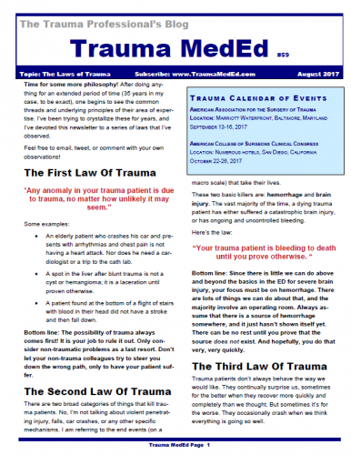
The August 2017 Trauma MedEd Newsletter Is Here!

Welcome to the current newsletter. In this one, I’ll be presenting and discussing some of the “Laws of Trauma” that I’ve observed over the years. I think you’ll find them interesting and amusing, and hopefully valuable. As a bonus, I’ll also include a copy of Norm McSwain’s Rules of Patient Care. Enjoy!

To download the current issue, just click here! Or copy this link into your browser: http://bit.ly/TME201708.

The “Egg Timer” Injury

Most patients with major traumatic injuries are handled in a very systematic way by both EMS and trauma centers. We have routines and protocols designed to provide rapid, quality care to these individuals. But over the years, I’ve begun to appreciate the fact that there is a very small subset of these patients who are different.

I term these patients as having an “egg timer injury”. These are patients who have only a certain number of minutes to live. This fact requires us to change the usual way we do things in order to save their lives or limbs. The usual routine may be too slow.

And unfortunately, no one can tell us exactly how many minutes are left on the timer. We only know that it’s ticking. Here are some examples of such  injuries:

  • Pericardial tamponade
  • Penetrating injury to the torso with profound hypotension
  • Orbital compartment syndrome

In each case, speed is of the essence. What can we do to decrease the time to definitive intervention? For prehospital providers, you may need to bypass a closer hospital that might not have the necessary resources at a particular time of day. Once at the hospital, the patient may need to bypass the emergency department and proceed straight to the OR. Or you may need to do a lateral canthotomy yourself, rather than waiting for an ophthalmologist to drive in only to have the patient lose their vision because of the  delay.

Bottom line: Remember that protocols are not necessarily etched in stone. They will cover 99.9% of cases you see. But that remaining 0.1%, the patients with the “egg timer injury”, will require you to think through what you know about the patient at the time, and make decisions about their care that may have a huge outcome on their life or livelihood. And as always, if you find that you must do things differently in the best interest of your patient, be sure to document what you knew and your thought processes thoroughly so you explain and/or justify your decision-making when you are invariably asked.

Syncope Workup in Trauma Patients – Updated With CPG

Syncope accounts for 1-2% of all ED visits, and is a factor in some patients with blunt trauma, especially the elderly. If syncope is suspected, a “syncope workup” is frequently ordered. Just what this consists of is poorly defined. Even less understood is how useful the syncope workup really is.

Researchers at Yale retrospectively looked at their experience doing syncope workups in trauma patients. They were interested in seeing what was typically ordered, if it was clinically useful, and if it impacted length of stay.

A total of 14% of trauma patients had syncope as a possible contributor to their injury. The investigators found that the following tests were typically ordered in these patients:

  • Carotid ultrasound (96%)
  • 2D Echo (96%)
  • Cardiac enzymes (81%)
  • Cardiology consult (23%)
  • Neurology consult (11%)
  • EEG (7%)
  • MRI (6%)

Most of this testing was normal. About 3% of cardiac enzymes were abnormal, as were 5% of carotid imaging and 4% of echocardiograms.

Important! Of the patients who underwent an intervention after workup, 69% could have been identified based on history, physical exam, or EKG and did not depend on any of the other diagnostic tests.

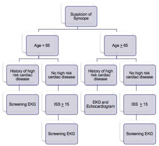
Is it possible to determine a subset of this population that may show a higher yield for this screening? Surgeons at Temple University in Philadelphia found that there was little utility in using carotid duplex studies. They did note that patients with a history of heart disease were more likely to have an abnormal EKG, and that an abnormal EKG predicted an abnormal echo. Overall, only patients with a history of significant cardiac comorbidity, older age, and higher ISS had findings requiring intervention.

Bottom line: Don’t just reflexively order a syncope workup when there is a question of this problem. Think about it first, because the majority of these studies are nonproductive. They are not needed routinely in trauma patients with “syncope” as a contributing factor.  Obtain a good cardiac history, and if indicated, order an EKG and go from there. See the practice guideline proposed by the Temple group below. And be sure to include the patients primary doctor in the loop!

References:

  1. Routine or protocol evaluation of trauma patients with suspected syncope is unnecessary. J Trauma 70(2):428-432, 2011.
  2. Syncope workup: Greater yield in select trauma population. Intl J Surg, accepted for publication June 27, 2017.