Pet Peeve: (Not So) Clever Medical Study Acronyms

I’m not a big fan of acronyms, although they do serve a purpose. We use them all the time providing medical care. CBC. CTA. CXR. ROSC. And a zillion others. And they can actually be helpful so you don’t have to say or write down some ridiculously long phrase. OMG.

But what really bothers me is the rise of researchers designing clever acronyms for medical studies. The first one , the University Group Diabetes Program (UGDP), was developed in the 1970s. It was actually shortened by journals and media to make for an easier presentation, not by the group themselves.

But then in the 1980s, the Multiple Risk Factor Intervention Trial (MRFIT) came along. It evaluated the impact of multiple interventions on cardiovascular mortality. Mr. Fit. Get it? This was the first of an ever growing number of studies that chose acronyms that were either cleverly related to the work in some way, or that made a catchy new word to help people remember it.

And the number of these acronyms has been growing rapidly. From 1992 to 2002, they increased from 245 to 4100, a 16-fold increase. There are now so many acronyms that many simple ones are being reused. And it seems like studies without an acronym are becoming the minority.

Plus, we’ve moved away from creating pure acronyms like UGDP that are derived from the first letter of each word. Now we use multiple letters from a word, skip some words altogether, or don’t even bother to use the words at all. There are MICHELANGELO, MATISSE, PICASSO, and EINSTEIN studies that were given the name just for the positive association. Nothing to do with the study at all.

This is all a warm-up for my next post, which reviews a geriatric trauma prognosis calculator from the PALLIATE consortium (Prognostic Assessment of Life and Limitations After Trauma in the Elderly). Groan! The title itself almost made me not want to read it. But I am compelled. Tune in Monday.

Reference: SearCh for humourIstic and Extravagant acroNyms and Thoroughly Inappropriate names For Important Clinical trials (SCIENTIFIC): qualitative and quantitative systematic study. BMJ. 2014;349:g7092.

Tips For Taking Care Of CSF Leaks

The management of CSF leaks after trauma remains somewhat controversial. The literature is sparse, and generally consists of observational studies. However, some general guidelines are supported by large numbers of retrospectively reviewed patients.

  • Ensure that the patient actually has a CSF leak. In most patients, this is obvious because they have clear fluid leaking from ear or nose that was not present preinjury. Here are the options when the diagnosis is less obvious (i.e. serosanguinous drainage):
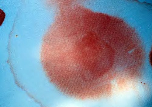
    • The “halo” or “double ring sign” is a form of pillow chromatography. The blood components separate from the CSF as they move through the pillow fabric, creating a clear ring or halo surrounding a bloody spot. This is the cheapest, fastest test and is actually fairly reliable.
    • High resolution images of the temporal bones and skull base. If an obvious breach is noted, especially if fluid is seen in the adjacent sinuses, then a CSF leak is extremely likely.
    • Glucose testing. CSF glucose is low compared to serum glucose.
    • Beta 2 transferrin assay. Don’t do it!! This marker is very specific to CSF. However, the test is expensive and results may take several days to a few weeks to receive. Most leaks will have closed before the results are available, making this a poor test.
  • Place the patient at bed rest with the head elevated. The basic concept is to decrease intracranial pressure, which in turn should decrease the rate of leakage. This same technique is used for management of mild ICP increases after head injury.
  • Consider prophylactic antibiotics carefully. The clinician must balance the likelihood of meningitis with the possibility of selecting resistant bacteria. If the likelihood of contamination is low and the patient is immunocompetent, antibiotics may not be needed.
  • Ear drops are probably not necessary. They may confuse the picture when gauging resolution of the CSF leak.
  • Wait. Most tramatic leaks will close spontaneously within 7-10 days. If it does not, a neurosurgeon or ENT surgeon should be consulted to consider surgical closure.

References:

  1. Brodie HA, Thompson TC. Management of complications from 820 temporal bone fractures. Am J Otol, 1997;18:188-197.
  2. Brodie HA. Prophylactic antibiotics for posttraumatic cerebrospinal fluid fistulas. Arch Otolaryngol Head, Neck Surg. 123:749-752.

Another Worthless Test? Serum / Urine Myoglobin

We often rely on diagnostic testing to augment our physical examination skills. These tests may be in the form of imaging that allows us to see things that we normally cannot, or measurements of body composition using laboratory testing.

If you look at the “menu” of tests that your hospital laboratory offers, it is very extensive. You can order just about any assay imaginable on any body fluid or tissue. Diagnosis of many of the clinical diseases or disorders that we treat has come to rely on some of these assays.

Let’s take rhabdomyolysis, for example. I’ve been writing about compartment syndrome in the last few posts. One of the byproducts of a full-blown compartment syndrome is muscle breakdown. Two of the well-known substances released from injured muscle are creatine kinase (CK) and myoglobin.

Many textbooks advise the clinician to monitor levels of these substances, since myoglobin is toxic to nephrons and may lead to kidney injury. So most trauma professionals routinely write orders for serial CK, myoglobin, as well as creatinine to monitor renal function.

But most clinicians do not know their laboratory as well as they think. Your lab has the capability to perform commonly requested tests rapidly and on site. But what about assays that are rarely ordered? Does it make sense to have the reagents necessary for these uncommon tests on hand at all times? They degrade over time, and may very well expire before they are ever used, costing money to replace.

So most hospital labs send uncommon assays out to larger labs that perform the test for a large geographic area. But how does the “send out” specimen get to that lab? By courier (if local) or more commonly, by delivery service (if remote). And obviously, this takes time. And some assays are complex and may take hours or days to perform.

At my hospital lab, a serum or urine myoglobin assay is a “send out” test. And if you ask, the lab will tell you that it takes 3-4 business days to get the result. So if you send it out Wednesday, you will have the result the following Monday! Does it make any sense to get serial myoglobins? Or even a single myoglobin test? By the time you get the result, your patient will be treated and gone!

Bottom line: Think about the tests that you order very carefully. If you are ordering something out of the ordinary, check with your lab. Is it a “send out” test? How long does it take to get a result? And more importantly, how expensive is it? These tend to be pricey due to the low frequency of processing.

Then do the math. Is it worth the wait and expense? Or can you get the same information using routine, in house testing? In the case of rhabdomyolysis, serum CK levels are good markers, as is visual inspection of the urine. If it’s any darker than a light yellow, there may be myoglobin present. A quick and dirty way to confirm is some inexpensive testing: a urine specimen that is dipstick positive for blood, and with microscopic analysis shows few if any RBCs usually means myoglobin. Voila! Diagnosis now, not in 4 days.

What The Heck? Final Answer: Progressive Back Pain After Heavy Lifting

In my last two posts, I described an athlete who developed significant pain in his lower back after rapidly escalating his weight lifting regimen. The pain was very localized to the paraspinal areas bilaterally, and serum CPK was elevated.

Congrats to Jay Slutsky for being the first to figure this one out. The suspected diagnosis was lumbar paraspinous muscle compartment syndrome. Compartment pressures were measured, and were found to be 78 and 26 mm Hg. A contrast MRI was obtained that showed swelling of both sets of paraspinal muscles.

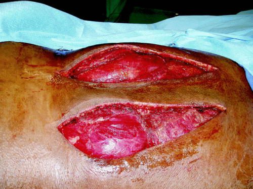
The patient was taken to the OR for fasciotomy.Source: Published paper

Note the bulging musculature above. Some areas appeared to be necrotic and did not bleed or contract. There were sharply debrided. The patient recovered quickly, with significant pain relief. The skin incisions were closed after several days, once swelling had subsided. He was well-healed and pain-free at his one month postop visit.

As you can see, any muscle surrounded by a rigid fascial compartment can develop a compartment syndrome. Typically, this requires direct trauma, but exertional compartment syndromes as in this case have been described in the legs of athletes as well. A history of a blow to the muscle group, or of very intense exercise should raise suspicion.

Physical findings of extreme pain that is very focal, coupled with discrete tenderness and firm muscle compartments, should confirm the potential diagnosis. Serum CPK is helpful for trending. Normal pressures in this muscle group tend to be in the single digits to low teens. They rise transiently during exercise, but usually return to normal shortly afterwards. “Normal” compartment pressures are not really known, so findings need to be coupled with CPK levels. Once the compartment pressure reaches the 30s, and especially if accompanied by high and rising CPKs, the syndrome is present. MRI is interesting, but not terribly helpful.

Treatment is typical for any compartment syndrome: release the muscle! A vertical incision centered over the bulging and tight muscle compartment is used. The wound is left open until swelling subsides enough to close the skin. Recovery is usually rapid, although some complain of a persistent low level of pain for a period of time. It is not known how soon these patients may resume sports or training.

Bottom line: Any patient with direct trauma or extreme exertion involving a muscle group is at risk for compartment syndrome. Physical exam, coupled with compartment pressure measurement if in doubt, are the mainstays of diagnosis. CPK levels may help in cases of uncertainty. As with any compartment syndrome, rapid diagnosis and fasciotomy is the key to preserving function and decreasing the likelihood of disability and chronic pain.

Related posts:

References:

  • Acute Exertional Lumbar Paraspinal Compartment Syndrome. Spine 35(25):E1529-E1533, 2010.
  • Lumbar paraspinal compartment syndrome. International Orthopaedics 36:1221-1227, 2011.
  • Paravertebral compartment syndrome after training causing severe back pain in an amateur rugby player: report of a rare case and review of the literature. BMC Musculoskelet Disord 14:259, 2013.


What The Heck? Part 2: Progressive Back Pain After Heavy Lifting

Yesterday, I described a case of a young athlete who developed progressive back pain after rapidly increased his deadlift weights. He presented to the hospital with back pain and inability to get up from a supine position. He had firm and tender paraspinal muscles in his lower back, but no other findings.

What to do next? Obviously, we need a bit more information on the bony structures. Other than run of the mill muscle strain, a compression fracture would be the next most common diagnosis. In this young, healthy athlete, a simple set of AP and lateral spine images should be sufficient. But if you opted for a CT scan, I won’t argue. In either case, the images were normal.

Since there is significant muscle pain and tenderness, a lab panel with a few extras is in order, as well. The usual electrolytes, etc were normal. Creatinine was 0.9, but CPK was 60,000!

Now what are you thinking? What’s the diagnosis, and what is the decision tree for treatment?

Add your comments below, or tweet them out. I’ll finish this topic up in the next post.