Closing Velocity And Injury Severity

Trauma professionals, both prehospital and in trauma centers, make a big deal about “closing velocity” when describing motor vehicle crashes.  How important is this?

So let me give you a little quiz to illustrate the concept:

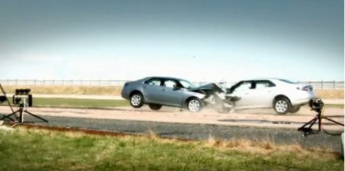
Two cars, of the same make and model, are both traveling on a two lane highway at 60 mph in opposite directions. Car A crosses the midline and strikes Car B head-on. This is the same as:

  1. Car A striking a wall at 120 mph
  2. Car B striking a wall at 60 mph
  3. Car A striking a wall at 30 mph

2010-saab-9-5-head-on-crash-test_100313384_m1

The closing velocity is calculated by adding the head-on components of both vehicles. Since the cars struck each other exactly head-on, this would be 60+60 = 120 mph. If the impact is angled there is a little trigonometry involved, which I will avoid in this example. And if there is a large difference in mass between the vehicles, there are some other calculation nuances as well.

So a closing velocity of 120 mph means that the injuries are worse than what you would expect from a car traveling at 60 mph, right?

Wrong!

In this example, since the masses are the same, each vehicle would come to a stop on impact because the masses are equal. This is equivalent to each vehicle striking a solid wall and decelerating from 60 mph to zero immediately. Hence, answer #2 is correct. If you remember your physics, momentum must be conserved, so both of these cars can’t have struck each other at the equivalent of 120 mph. The injuries sustained by any passengers will be those expected in a 60 mph crash.

If you change the scenario a little so that a car and a freight train are traveling toward each other at 60 mph each, the closing velocity is still 120 mph. However, due the the fact that the car’s mass is negligible compared to the train, it will strike the train, decelerate to 0, then accelerate to -60 mph in mere moments. The train will not slow down a bit. For occupants of the car, this would be equivalent to striking an immovable wall at 120 mph. The injuries will probably be immediately fatal for all.

Bottom line: Closing velocity has little relationship to the injuries sustained for most passenger vehicle crashes. Those injuries will be consistent with the speed of the vehicle the occupants were riding, and not the sum of the velocities of the vehicles. 

Why Is My Trauma Patient On Oxygen?

How many times has this happened to you? You walk into a young, healthy trauma patient’s room and discover that they have nasal prongs and oxygen in place. Or better yet, these items appear overnight on a patient who never needed them previously. And the reason? The pulse oximeter reading had been “low” at some point.

loc-0424-cs-belding-pet-masks-11-1024x6821

This phenomenon of treating numbers without forethought has been one of my pet peeves for years. Somehow, it is assumed that an oximetry value less than the standard “normal” requires therapy. This is not the case.

In young, healthy people the peripheral oxygen saturation values (O2 sat) are typically 96-100% on room air. As we age, the normal values slowly decline. If we abuse ourselves (smoking, working in toxic environments, etc), lung damage occurs and the values can be significantly lower. Patients with obstructive sleep apnea will have much lower numbers intermittently through the night.

So when does a trauma inpatient actually need supplemental oxygen? Unfortunately, the literature provides little guidance on what “normal” really is in older or less healthy patients. Probably because there is no norm. The key is that the patient must need oxygen therapy.

But how can you tell? Examine them! Talk to them! If the only abnormal finding is patient annoyance due to the persistent beeping of the machine, they don’t need oxygen. If they feel anxious, short of breath, or have new onset tachycardia, they probably do. Saturations in the low 90s or even upper 80s can be normal for the elderly and smokers.

Bottom line: Don’t get into the habit of treating numbers without thinking about them. There are lots of reasons for the oximeter to read artificially low. There are also many reasons for patients to have a low O2 sat reading which is not physiologically significant. So listen, talk, touch and observe. Set the alarm level to 90%, or even lower. And if your patient is comfortable and has no idea that their O2 sat is low, turn off the oxygen and toss the oximeter out the window.

The Cribari Grid And Over/Undertriage

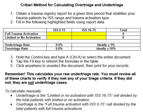
I’ve spent some time discussing undertriage and overtriage. I frequently get questions on the “Cribari grid” or “Cribari method” for calculating these numbers. Dr. Cribari is a previous chair of the Verification Review Subcommittee of the ACS Committee on Trauma. He developed a table-format grid that provides a simplified method for calculating these numbers.

But remember, the gold standard for calculating over- and undertriage is examining each admission to see if they met any of your trauma activation triage criteria. The Cribari method is designed for those programs that do not check these on every admission. It is a surrogate that allows you to identify patients with higher ISS that might have benefited from a trauma activation.

So if you use the Cribari method, use it as a first pass to identify potential undertriage. Then, examine the chart of every patient in the undertriage list to see if they meet any of your activation criteria. If not, they were probably not undertriaged. However, you must then look at their injuries and overall condition to see if they might have been better cared for by your trauma team. If so, perhaps you need to add a new activation criterion. And then count that patient as undertriage, of course.

I’ve simplified the calculation process even more and provided a Microsoft Word document that automates the task for you. Just download the file, fill in four values in the table, update the formulas and voila, you’ve got your numbers! Instructions for manual calculations are also included. Download it by clicking the image below or the link at the end of this post.

cribarigrid

Download the calculator by clicking here

Related posts:

Chest Tube Repositioning – Final Answer

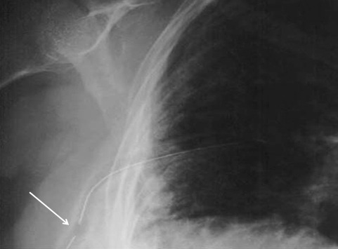
So you’re faced with a chest tube that “someone else” inserted, and the followup chest xray shows that the last drain hole is outside the chest. What to do?

Well, as I mentioned, there is very little written on this topic, just dogma. So here are some practical tips on avoiding or fixing this problem:

  • Don’t let it happen to you! When inserting the tube, make sure that it’s done right! I don’t recommend making large skin incisions just to inspect your work. Most tubes can be inserted through a 2cm incision, but you can’t see into the depths of the wound. There are two tricks:
    • In adults with a reasonable BMI, the last hole is in when the tube markings show 12cm (bigger people need bigger numbers, though)
    • After insertion, get into the habit of running a finger down the radiopaque stripe on the tube all the way to the chest wall. If you don’t feel a hole (which is punched through the stripe), this will confirm that the it is inside, and that the tube actually goes into the chest. You may laugh, but I’ve seen them placed under the scapula. This even looks normal on chest xray!
  • Patients with a high BMI may not need anything done. The soft tissue will probably keep the hole occluded. If there is no air leak, just watch it.
  • If the tube was just put in and the wound has just been prepped and dressed, and the hole is barely outside the rib line, you might consider repositioning it a centimeter or two. Infection is a real concern, so if in doubt, go to the next step.
  • Replace the tube, using a new site. Yes, it’s a nuisance and requires more anesthetic and possibly sedation, but it’s better than treating an empyema in a few days.

Related posts:

Chest Tube Repositioning – Part 2

Yesterday I presented the problem of the malpositioned chest tube, specifically one that is not completely in the pleural space. This one is way out:

So what do the doctor books say? Well, the first thing you will discover if you try to look it up is that THERE IS NO LITERATURE ON THIS COMMON PROBLEM! There are a few papers on tubes placed in the fissure and tubes inserted into the lung parenchyma. But there are only a few mentions of tubes with holes still outside the chest.

I’ve gotten a number of comments, including “you can push them in a little”, “take it out and put in another”, and “never push them in.” Since we don’t have any science to guide us, we have to use common sense. But remember, I’ve shown you plenty of examples where something seems reasonable, but turns out to be ineffective or downright harmful.

There are three principles that guide me when I face this problem:

  • Prevention is preferable to intervention
  • Do no (or as little as possible) further harm
  • Be creative

Tomorrow, I’ll finish this series and provide some tips and guidelines to help manage this problem using the principles outlined above.