Does Aspirin Add Anything To DVT Prophylaxis?

Venous thromboembolism (VTE) is an ongoing problem for trauma professionals. Most trauma programs have settled on their own flavor of screening, prophylaxis, and treatment once the problem actually surfaces in a patient. Most prophylaxis centers around a combination of mechanical (leg squeezers) and chemical (some type of heparin) management.

Aspirin has been used for prophylaxis for elective orthopedic surgery, and occasionally in trauma patients managed by orthopedic surgeons for years. Existing literature supporting this has been sparse and unconvincing. But since VTE involves platelets as part of the process, why not have another look?

A recently published paper from Scripps in San Diego looked tried to gauge the effect of aspirin on trauma patients where taking it before they were injured. Novel idea. Can the findings be useful? The authors performed a retrospective, case-controlled study of patients who developed post-traumatic deep venous thrombosis (DVT). The patients were matched for 7 covariates, and the authors looked at an additional 26 risk factors. Those taking aspirin pre-injury were compared with those who were not.

Here are the factoids:

  • 172 cases were identified over the 5 ½ year study, and 62 (36%) were excluded because a matched control could not be found
  • 7% of the remaining110

    patients were taking aspirin (why?)

  • 13% of controls were taking aspirin
  • 7% were taking warfarin, and 4% were taking clopidogrel
  • The mean age was 52, ISS was 13-14, and hospital stay was 7-10 days (!)
  • Multivariate analysis showed a significant protective effect from DVT with a risk ratio of 0.17 (!!)
  • But this effect was found only when used in conjunction with heparin prophylaxis after admission

Bottom line: Interesting findings. What does it mean? First, this is a very small retrospective study. It was conducted over 5+ years, so changes in VTE screening and prophylaxis may have occurred at this hospital. But even so, the finding were compelling. The biggest problem is that we can’t expect people to predict that they will need to start taking aspirin. But the study does raise the interesting question of whether it might be helpful to start taking it as soon as the patient arrives at the hospital. This is one of those thought provoking studies that should prompt someone (hint hint) to design a nice prospective study to see if this ultra-cheap drug might help us bring down our VTE rates even more.

Reference: Aspirin as added prophylaxis for deep vein thrombosis in trauma: A retrospective case-control study. J Trauma 80(4):625-630, 2016.

Potentially Avoidable Pediatric Transfers

Pediatric emergency and trauma care is not readily available across a sizable chunk of the US, particularly in rural areas. Couple this with the fact that many rural emergency providers are not necessarily trained in emergency medicine and may have little recent pediatric training fosters the common practice of transferring these injured children to a higher level of care.

And unfortunately, many of these transferred children have relatively simple issues that really don’t actually need a transfer. Some studies have reported that up to 40% of children sent to tertiary pediatric centers are sent home in less than 24 hours.

Most research in this area focuses on single medical center experiences. An article currently in press looks at the experience of the entire state of Iowa over a 10 year period. The authors looked at all claims data for children between ages 8 days and 18 years. Children who were transferred were compared to those who were not.

Here are the factoids:

  • 2 million cases were included in the study, and only 1% were transferred (21,319)
  • Children in rural areas were transferred 3x more often than those in urban areas
  • Only 63% were transferred to a designated children’s hospital, and 45% were sent to an ED rather than direct transfer to an inpatient bed
  • 39% were potentially avoidable transfers, meaning that they were discharged from the receiving ED or the hospital within 24 hours of admission
  • Two of the top 5 reasons for transfer were trauma related: fracture, and TBI without blood in the head.
  • The cost for potentially avoidable transfers in the top 5 categories was $2 million dollars (!)

Bottom line: This is a very comprehensive study that shows the magnitude and cost consequences of potentially inappropriate pediatric transfers. It was not designed to figure out what to do about it, but it provides some insight for the problem solvers out there. Since we know the top 5 transfer diagnoses (seizure, fracture, TBI without bleeding, respiratory infection, and asthma), we can start to work on systems to provide education to rural providers on these topics, as well as real-time interaction to help them determine the 60% that really do need a higher level of care. Telemedicine will eventually be a big part of this, but most areas around the country are still struggling to figure out the details. Stay tuned!

Reference: Potentially Avoidable Pediatric Interfacility Transfer is a Costly Burden for Rural Families: A Cohort Study. Acad Emerg Med 28 March 2016, in press.

A Scan That Can Find Clots Anywhere In The Body

Our current technology for identifying venous thromboembolism (VTE) / deep venous thrombosis (DVT) consists of duplex ultrasonography, and sometimes, CT angiography. Both are relatively noninvasive and painless (unlike the old-fashioned venography of days gone by.

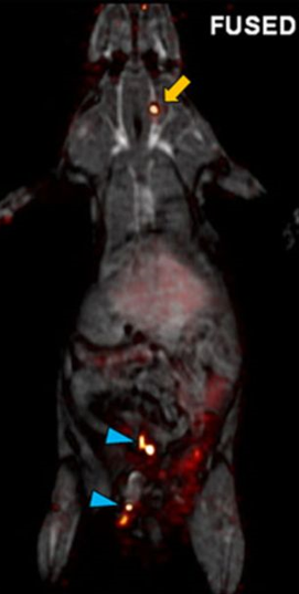
Researchers at the Massachusetts General Hospital have been working with different chemical probes that could adhere to clot and allow it to be identified on a PET scan. After experimenting with a number of fibrin-targeting peptides they settled upon one called copper fibrin-binding peptide 8 (Cu-FBP8). It was found to have a high affinity for clot, remain stable, and clear quickly from the animal.

A series of rats were subject to a surgically induced thrombus in the carotid or femoral arteries, or a sham operation. The animals were then imaged by CT/PET scan after injection with the Cu-FBP8 probe. The authors found that the probe worked as expected, identifying clot immediately. They were also able to follow resolution over the days following induction. 

Here is a whole-body fused CT/PET scan of one of the animals with both carotid artery (yellow arrow) and femoral artery (blue arrows) clot.

Bottom line: This is a potentially exciting tool that could make it much easier for us to identify DVT and VTE. It could also help us understand the etiology and incidence of PE as well. But as with all animal studies, it remains to be seen whether this will translate into a useful test for humans. Stay tuned, as it will probably take about 3 years to find out the answer.

Reference: 

Multisite Thrombus Imaging and Fibrin Content Estimation With a Single Whole-Body PET Scan in Rats. Arterioscler, Thromb, Vasc Biol 35(10):2114-2121, 2015.

Pulmonary Embolism and DVT in Trauma

We have long assumed that pulmonary emboli start as clots in the deep veins of the legs (or pelvis), then break off and float into the branches of the pulmonary artery in the lungs. A huge industry has developed around how best to deal with or prevent this problem, including mechanical devices (sequential compression devices), chemical prophylaxis (heparin products), and physical devices (IVC filters).

The really interesting thing is that less than half of patients who are diagnosed with a pulmonary embolism have identifiable clots in their leg veins. In one study, 26 of 200 patients developed DVT and 4 had a PE. However, none of the DVT patients developed an embolism, and none of the embolism patients had a DVT! How can this kind of disparity be explained?

Researchers at the Massachusetts General Hospital retrospectively looked at the correlation between DVT and PE in trauma patients over a 3 year period. DVT was screened for on a weekly basis by duplex venous ultrsonagraphy. PE was diagnoses exclusively using CT scan of the chest, but also included the pelvic and leg veins to look for a source. A total of 247 patients underwent the CT study for PE and were included in the study.

Here are the factoids:

  • Forty six patients had PE (39% central, 61% peripheral pulmonary arterial branches) and 18 had DVT (16 seen on the PE CT and 2 found by duplex)
  • Of the 46 patients with PE, only 15% had DVT
  • All patient groups were similar with respect to injuries, injury severity, sex, anticoagulation and lengths of stay
  • Interestingly, 71% of PE patients with DVT had a central PE, but only 33% of patients without DVT had a central PE.

The authors propose 4 possible explanations for their findings:

  1. The diagnostics tools for detecting DVT are not very good. FALSE: CT evaluation is probably the “gold standard”, since venography has long since been abandoned
  2. Many clots originate in the upper extremities. FALSE: most centers do not detect many DVTs in the arms
  3. Leg clots do not break off to throw a PE, they dislodge cleanly and completely. FALSE: cadaver studies have not shown this to be true
  4. Some clots may form on their own in the pulmonary artery due to endothelial inflammation or other unknown mechanisms. POSSIBLE

An invited critique scrutinizes the study’s use of diagnostics and the lack of hard evidence of clot formation in the lungs.

Bottom line: this is a very intriguing study that questions our assumptions about deep venous thrombosis and pulmonary embolism. More work will be done on this question, and I think the result will be a radical change in our use of anticoagulation and IVC filters over the next 3-5 years.

Reference: Pulmonary embolism and deep venous thrombosis in trauma: are they related? Arch Surg 144(10):928-932, 2009.

More On CT Usage In Unstable Patients

Yes, it is practically dogma that CT should not be used in unstable trauma patients. Either they go directly to the OR, or an attempt to stabilize them is briefly undertaken in the trauma bay. And as you know, I’m not a big believer in dogma. But this one has withstood the test of time. You can see my comments about a previous paper below in the related posts.

But now some authors in Colombia have published a paper that seems to call this idea into question. Could it be true? Read carefully!

This was a small, retrospective review of patients from a large Level I government designated trauma center. They reviewed their experience over a two year period, identifying all hemodynamically unstable patients in the registry. They excluded dead patients, those with isolated head injury, and any who had surgery at an outside hospital prior to transfer.

Here are the factoids:

  • 171 patients were reviewed, and of course they tended to be young males
  • 91 went straight to the OR, and 80 were taken to CT first
  • “Unstable” patients were defined as having SBP < 100 and/or HR > 100
  • Mechanism of injury for the OR group was 95% penetrating, but for the CT group was about 50:50 penetrating/blunt
  • The mean SBP and HR for the “unstable” patients taken to CT were 92 and 110, respectively
  • Mortality was the same for both groups (18% OR vs `13% CT)

Bottom line: The authors concluded that it is permissible to take unstable patients to CT if you don’t spend too much time there based on similar mortality rates. But the problem was that I don’t consider their patients to have been unstable! Mean SBP in their “unstable” group was over 90 torr and the heart rate was only 110! The lowest SBP was only 79. And mortality is too crude of an outcome to rely on. Furthermore, the patients they took to CT tended to have blunt mechanisms, and may not have had ample efforts at resuscitation in the trauma bay first, or may have met criteria to go to CT anyway (see related posts below).

Reference: Computed tomography in hemodynamically unstable severely injured blunt and penetrating trauma patients. J Trauma 80(4):597-603, 2016.