Liver Laceration And Liver Function Tests

Over the years I’ve seen a number of trauma professionals, both surgeons and emergency physicians, order liver transaminases (SGOT, SGPT) and bilirubin in patients with liver laceration. I’ve never been clear on why, so I decided to check it out. As it turns out, this is another one of those “old habits die hard” phenomena.

Liver lacerations, by definition, are disruptions of the liver parenchyma. Liver tissue and bile ducts of various size are both injured. Is it reasonable to expect that liver function tests would be elevated? A review of the literature follows the typical pattern. Old studies with very few patients.

From personal hands-on observations, the liver tissue itself tears easily, but the ducts are a lot tougher. It is fairly common to see small, intact ducts bridging small tears in the substance of the liver. However, larger injuries can certainly disrupt major ducts, leading to major problems. But I’ve never seen obstructive problems develop from this injury.

A number of papers (very small, retrospective series) have shown that transaminases can rise with liver laceration. However, they do not rise reliably enough to be a good predictor of either having an injury, or the degree of injury. Similarly, bilirubin can be elevated, but usually not as a direct result of the injury. The most common causes are breakdown of transfused or extravasated blood, or from critical care issues like sepsis, infection, and shock.

Bottom line: Don’t bother to get liver function tests in patients with known or suspected injury. Only a CT scan can help you find and/or grade the injury. And never blame an elevated bilirubin on the injury. Start searching for other causes, because they will end up being much more clinically significant.

References:

  • Evaluation of liver function tests in screening for intra-abdominal injuries. Ann Emerg Med 20(8):838-841, 1991.
  • Markers for occult liver injury in cases of physical abuse in children. Pediatrics 89(2):274-278.
  • Combination of white blood cell count with liver enzymes in the diagnosis of blunt liver laceration. Am J Emerg Med 28(9):1024-1029, 2010.

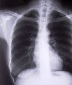
Do You Really Need To Repeat That Trauma Bay Xray?

It happens all the time. You get that initial chest and/or pelvic xray in the resuscitation room while evaluating a blunt trauma patient. A few minutes later the tech returns with another armful of xray plates to repeat them. Why? The patient was not centered properly and part of the image is clipped.

Where is the left side of the chest, and do we care?

Do you really need to go through the process of setting up again, moving the xray unit in, watching people run out of the room (if they are not wearing lead, and see my post below about how much radiation they are really exposed to), and shooting another image? The answer to the question lies in what you are looking for. Let’s address the two most common (and really the only necessary) images needed during early resuscitation of blunt trauma.

First, the chest xray. You are really looking for 3 things:

  • Big air (pneumothorax)
  • Big blood (hemothorax)
  • Big mediastinum (hinting at aortic injury)

Look at the clipped xray above. A portion of the left chest wall is off the image. If there were a large pneumothorax on the left, would you be able to see it? What about a large hemothorax? And the mediastinum is fully included, so no problem there. So in this case, no need to repeat immediately.

The same thing goes for the pelvis. You are looking for gross disruption of the pelvic ring, especially posteriorly because this will cause you to intervene in the ED (order blood, consider wrapping the pelvis). So if parts of the edges or top and bottom are clipped, no big deal.

Bottom line: Don’t let the xray tech disrupt the team again by reflexively repeating images that are not technically perfect. See if you can use what you already have.  And how do you decide if you need to repeat it later, if at all? Consider the mechanism of injury and the physical exam. Then ask yourself if there is anything you could possibly see that was not imaged the first time that would change your management in any way. If not, you don’t need it. But it certainly will irritate the radiologists!

The Impact Of Radiographic Image Sharing Systems On Trauma Transfers

There has been a big push to implement systems of trauma centers across the US, primarily at the state level. This move to get the right patient to the right hospital has resulted in an increased number of transfers, and rightly so. However, the referring hospital frequently performs some radiographic imaging before transfer.

So it is critical that both the patient and their imaging get to the receiving hospital for good continuity of care. Failure to do so results in re-imaging, additional exposure to radiation, delays in care, and potentially increased costs. Radiologists may be reluctant to read outside images because they generally will not get paid for it.

Unfortunately, there are lots of barriers to getting those images to the receiving trauma center. They may forget to send a disc. The disc may not work on the receiving hospital’s computers. A direct connection between PACS systems may be lacking, or may not work. In any case, patient care may suffer.

Cloud solutions using web-based software and an intermediary for image storage and delivery have been around for years. Their use is inconsistent around the US, mainly because they cost money. A group in Ohio looked at the impact of implementing one of these system on the incidence of cost of re-imaging at their Level I trauma center. Four years of patient transfer data were reviewed for imaging at the first hospital, re-imaging at the trauma center, and charges. The authors compared re-imaging rates before and after the availability of the cloud sharing system.

Here are the factoids:

  • 1,081 transfers occurred during the study period, and 639 (59%) had at least one CT prior to transfer
  • 345 repeat scans were performed on 222 patients (35%)
  • The most common repeats were head CT (32%) and cervical spine (23%)
  • The overall re-scan rate was significantly higher before the cloud service was available (38%) vs after (28%)
  • If patient data was available from the cloud service, the re-scan rate dropped to 23% (??!)
  • Mean hospital charges for re-CT dropped from $1046 to $589

Bottom line: This study is interesting, but could use some improvement. It is older data (2009-2012), from the early days of these cloud services. Centers were a little less facile using them, which may have contributed to some of the soft numbers above. And the use of charge data rather than costs is old-school. 

Re-scanning a quarter of the patients, even when cloud images were available, is just not acceptable. However, this paper does suggest that there are real benefits, as re-scan rates and (presumably) costs should decrease. Radiation exposure would definitely drop, too.

The key to making a cloud sharing system work, or any other system for that matter (VPN, optical discs, etc), is to make it part of your PI program. Every transfer in needs to be scrutinized, and if an image transfer issue is found, quick feedback to the referring hospital needs to occur to ensure that it doesn’t happen again.

Reference: Implementation of an image sharing system significantly reduced repeat computed tomographic imaging in a regional trauma system. J Trauma 80(1):51-56, 2016.

May Trauma MedEd Newsletter Is Coming!

The May issue of Trauma MedEd is coming soon! The topic? What everyone loves to hate! The electronic Trauma Flow Sheet. 

Here is what you will find inside:

  • Why the electronic trauma flow sheet (TFS)?
  • Human factors
  • Machine shortcomings
  • What to do if you’ve already switched?
  • The real bottom line
  • And what next?

Subscribers will receive it next weekend; everyone else will have to wait another week.

Subscribe now and be sure to get it first!  So sign up for early delivery now by clicking here!

Pick up back issues here!

Where Do Pulmonary Emboli Really Come From?

For a long time, we “knew” that pulmonary emboli were a possible and dreaded complication of deep venous thrombosis (DVT). However, we are beginning to discover that this is not always the case. The group in San Diego decided to see if there really are two different types of PE in trauma, and what that means.

Here’s another VTE paper from Scripps Mercy Hospital, a level I trauma center in San Diego. It looked at 5 ½ years of their experience with adult trauma patients who were routinely screened for DVT. Any of these patients who developed a PE within 6 weeks of admission were evaluated further.

Here are the factoids:

  • Duplex screening from groin to ankle was carried out twice weekly in ICU patients, and once weekly in ward patients
  • Surveillance was carried out if the patient would be non-ambulatory for more than 72 hours, or were at moderate or higher risk for DVT using the ACCP guidelines
  • Nearly 12,000 patients were evaluated by the trauma service and 2,881 underwent surveillance
  • 31 patients (1%) developed a PE
  • 12 of these 31 had DVT identified before or immediately after their PE. Clot was below-knee in 9 (!), above-knee in 2, and in the IJ in one.
  • 19 patients had PE but no DVT identified (de novo PE, DNPE)
  • DNPE tended to be single and peripherally located, and associated with rib fractures, pulmonary contusions, blood transfusions, and pneumonia
  • DVT + PE were more often found in multiple lobes or bilaterally

Bottom line: Like most, this is not a perfect study, but it’s a really good one. It is looking more and more likely that some PEs arise de novo, without any associated DVT. These clots are more likely to be linked to some type of inflammatory process, and have a tendency toward causing more of the classic signs and symptoms of PE. There are still lots of questions to be answered, like do you need to anticoagulate the de novo PEs? But for now, no change in practice. Just be aware that these might not be as bad as they seem.

Reference: Pulmonary embolism without deep venous thrombosis: de novo or missed deep venous thrombosis? J Trauma 76(5):1270-1281, 2014.