What The Heck? Final Answer

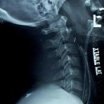
This one was a bit tricky. I chose it because it looks like there is an extra tube in the neck. You can see two stripes traveling from the mouth down the neck. The one closest to the cervical spine is in the esophagus, an orogastric tube. The other one passes anterior to it, in the trachea, so it is the orotracheal tube. But what about the tube shaped density that is located in the posterior pharynx that looks like it is angled forward toward the trachea? Did someone lose something?

If you think about it though, you should conclude it’s something weird. There is no radiopaque stripe on it, which rules out most common tubes. The only thing of that size and shape that comes to mind is a nasopharyngeal airway tube. However, these have a flange on the nasal end, so it couldn’t just pass inwards through the nose. And who in their right mind would put it in the mouth to be swallowed? Plus, the orientation of it is unusual, heading forward toward the trachea.

You have to look at the rest of the clues on the radiograph. It’s easy to get suckered if you just focus on the obvious. What are those objects located between the two tube stripes in front of C6? Surgical clips. What are those O-shaped objects at the angle of the mandible that disappear behind the XTABLE LAT marker? Surgical skin closure staples.

So this is a postoperative patient. If you follow the object, it actually moves toward the skin, and beyond! This patient was stabbed in the neck and underwent a surgical exploration with control of bleeding. A surgical drain was placed due to concern for leakage from the pharynx or salivary glands. The drain actually leaves the side of the neck, just anterior to the sternocleidomastoid muscle.

Remember to look at everything on a radiograph, especially if you don’t have the clinical story behind it. The eye normally focuses on the obvious, leading the viewer to make assumptions based on their expectations. This can easily get you in trouble, so beware! And don’t forget that you are looking at a 2-D image, so there is no way to tell where any object is in the third dimension. It may be in the front, the back, or under the patient in their clothing!

When To Image The Aorta In Blunt Trauma

Blunt injury to the thoracic aorta is one of those potentially devastating ones that you (and your patient) can’t afford to miss. Quite a bit has been written about the findings and mechanisms. But how do you put it all together and decide when to order a screening CT?

There are a number of high risk findings associated with blunt aortic injury. Recognize that they are associated with the injury, but are still not very common. They are:

  • Fractures of the sternum or first rib
  • Wide mediastinum
  • Displacements of mediastinal structures (left mainstem down, trachea right, esophagus right)
  • Loss of the aortopulmonary window
  • Apical cap over the left lung

Here’s a sensible method for screening for blunt aortic injury, using CT scan:

  • Reasonable mechanism (fall from greater than 20 feet, pedestrian struck, motorcycle crash, car crash at “highway speed”) PLUS any one of the high risk findings above.
  • Extreme mechanism alone (e.g. car crash with closing velocity at greater than highway speed, torso crush)

Note on torso crush: I have seen three aortic injuries from torso crush in my career, one from a load of plywood falling onto the patient’s chest, one from dirt crushing someone when the trench they were digging collapsed, and one whose chest was run over by a car.

Related post:

Technology: EEG Monitoring Using A Smartphone App

Remember when EEG monitoring in patients with severe TBI looked like a maze of multicolored spaghetti plugged into a small refrigerator? Well, technology is advancing rapidly and the hardware is shrinking fast.

This EEG monitor uses an EEG headset, which has fewer leads than the old standard. The headset connects to a Nokia smartphone using a wireless connection. And while it can’t compete with a regular EEG on fine detail like localizing seizure foci, it should easily be able to measure something as crude as burst suppression in trauma patients in pentobarb coma.

EEG headset

Expect more advances like this. Computing and monitoring is leaving the realm of the dedicated (and physically large) device, and moving toward handheld monitoring using off-the-shelf hardware like smartphones.