Predicting Bleeding In Patients With Stable Pelvic Fractures

Bleeding is a well-recognized complication of severe pelvic fracture. Certain fracture patterns, usually with significant involvement of the posterior portions of the ring, are associated with significant bleeding. Most of these fractures are unstable to some degree.

Stable pelvic fractures (those that do not require internal or external fixation) are not generally prone to a large amount of bleeding. However, it can occur on occasion, and surgeons at the Massachusetts General Hospital have devised a simple prediction system so patients more likely to bleed can be identified and monitored more closely.

They retrospectively looked at their stable pelvic fracture population over 5+ years. A total of 391 patients with stable pelvic injury were identified. Of those, 280 never required transfusion and 111 did. Of the latter, only 15 bled from their stable pelvic fractures. 

The authors found the following three significant indicators of bleeding from stable pelvic fractures:

  • Admission hematocrit < 30%
  • Pelvic hematoma on CT
  • Any systolic blood pressure < 90 mm Hg

Bottom line: This is a simple, retrospective study with low numbers. However, the three indicators commonly indicate significant early bleeding in any trauma patient, so it makes sense to apply it here, too. If a patient meets one or two criteria, consider monitoring in the ICU and consider angiography. If all three or met, strongly consider appropriate intervention (angiography if good blood pressures can be maintained, or fixation and/or preperitoneal packing if not).

Reference: Predictors of bleeding from stable pelvic fractures. Arch Surg 146(4):407-411, 2010.

Get Microsoft Silverlight

Technology: A New Teaching Tool For Orthopedic Injury

Here’s a look at some new technology (made from five pieces of old technology) to help injured patients follow their activity and exercise regimens better after injury. It allows patients to “look beneath the surface” of their injured extremity to get a better idea of what is wrong and why they need to be compliant to heal.

Microsoft Research Labs cobbled together a projection unit from a handheld projector, a digital camera and an infrared camera. The control unit consists of a wireless controller and a laser pointer. Put them together and you can superimpose stock injury images over a patient’s extremity, or review images on a wall.

Two physical therapists did an uncontrolled test on several patients and indicated that overall compliance with the therapy regimen seemed to be better. Obviously, this is not sound science. But it does have some potential in allow physicians and therapists to give a better explanation about what is injured and what needs to be done about it. In my opinion, this could be generalized to just about any internal injury, and can provide an easy to understand teaching tool for trauma professionals.

Anatomic injury projector

How To Troubleshoot Air Leaks in Chest Tube Systems

An air leak is a sure-fire reason to keep a chest tube in place. Fortunately, many air leaks are not from the patient’s chest, but from a plumbing problem. Here’s how to locate the leak.

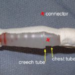
To quickly localize the problem, take a sizable clamp (no mosquito clamps, please) and place it on the chest tube between the patient’s chest and the plastic connector that leads to the collection system. Watch the water seal chamber of the system as you do this. If the leak stops, it is coming from the patient or leaking in from the chest wall.

If the leak persists, clamp the soft Creech tubing between the plastic connector and the collection system itself. If the leak stops now, the connector is loose.

If it is still leaking, then the collection system is bad or has been knocked over.

Here are the remedies for each problem area:

  • Patient – Take the dressing down and look at the skin entry site. Does it gape, or is their obvious air hissing and entering the chest? If so, plug it with petrolatum gauze. If not, the air is actually coming out of your patient and you must wait it out.
  • Connector – Secure it with Ty-Rap fasteners or tape (see picture). This is a common problem area.
  • Collection system – The one-way valve system is not functioning, or the system has been knocked over. Click here for an example. Replace it immediately.

Note: If you are using a “dry seal” system (click here for more on this) you will not be able to tell if you have a leak until you fill the seal chamber with some water.

Medication Alert! Dabigatran and Head Trauma

First, there was warfarin, a cheap and effective way of treating deep venous thrombosis (DVT) and pulmonary embolism (PE) in trauma patients. Unfortunately, there is plenty of literature that shows the added risk that this drug poses in injured patients, particularly in head injury. Because of this, many trauma centers have developed “rapid reversal protocols” to quickly restore vitamin K dependent clotting factors in an attempt to improve outcomes. To see our protocol, click here.

Next came clopidogrel (Plavix), which is used to prevent clotting in vascular disease. It irreversibly inhibits platelet aggregation. Counteracting this drug is more complicated due to its long half-life. Platelet infusions are required, but the infused platelets are inhibited by any remaining drug in the plasma. This requires the use of lots of platelets to get some meaningful clot to form again.

Now, we have direct thrombin inhibitors (DTI). Hirudins were the first used, and were never an issue in trauma patients. And their short half-lives obviate the need for reversal. The newest DTIs (argatroban and dabigatran) are a real problem in trauma. Argatroban is not a problem, because it is given by IV only. But dabigatran (Pradaxa) has just been approved for oral use within the last year.

According to the package insert, “there is no antidote to dabigatran etexilate or dabigatran.” And also “dabigatran can be dialyzed (protein binding is low), with the removal of about 60% of drug over 2 to 3 hours; however, data supporting this approach are limited.”

We will be seeing patients taking this drug in the near future. What do we do if they are trauma victims with bleeding in critical places, like the brain? At Regions, we have developed a proposed guideline that combines oral charcoal, dialysis, transfusions and optionally, activated Factor VII. Click here to download the protocol.

If anyone has any experience with these patients, please comment below. And everyone else, keep your fingers crossed!

Related posts:

Protocols:

Thanks to Colleen Morton MD for developing the dabigatran reversal protocol

DPL: A Dying Art?

Diagnostic peritoneal lavage (DPL) was invented by David Root at this hospital (Ancker Hospital, which then became St. Paul Ramsey, now Regions Hospital) in the 1960’s. It enjoyed its heyday during the 70’s and 80’s, when it was done hundreds of times per year at most major trauma hospitals. Now, we do it about 5 times per year. What happened?

As you know, DPL is a qualitative test. It gives a yes/no answer to the question “does this patient need an operation?” based on red and white blood cell counts. During the mid-1980s, CT scanning was introduced, which provides much more quantitative information about injuries in the abdominal cavity. The improved ability to diagnose abdominal injury, especially solid organ injury, has led to the demise of DPL.

Most solid organ injury results in some free blood in the peritoneal cavity. It doesn’t take much blood (10 cc of whole blood mixed with 1 liter of infused crystalloid) to exceed the threshold of 100,000 RBC per ml of aspirate that will send the surgeon off to the OR. Therefore, pretty much any liver or spleen laceration would have to be taken to the OR based on a DPL.

But we know that very few liver/spleen injuries actually need an operation. So DPL cannot be used, or the negative laparotomy rate for blunt trauma would escalate. The other downside to DPL is that it’s not possible to get all of the infused crystalloid back out of the abdomen. This leads to a confusing amount of free fluid seen on any CT scan done after a DPL.

So DPL is now down but not out. Some practical pointers:

  • DPL should be used primarily as a backup to an equivocal or unbelievable FAST exam in an unstable patient. An example would be a patient who is hypotensive, has a negative FAST and no other obvious bleeding sources.
  • Remember to insert a gastric tube and urinary catheter so the stomach and bladder are decompressed before the procedure. The easiest way to remember this is to tape these catheters to the DPL procedure tray.
  • A DPL is actually 2 procedures: peritoneal tap and lavage. Once the catheter is in, it should be aspirated. If 10cc of gross blood is returned, the test is positive and the patient needs to go immediately to OR.
  • For blunt trauma, the threshold for RBC per µl is 100,000. The threshold for WBC is 500 per µl. If particulate material or weird colors are seen (stool or bile), the test is also considered positive. Send the sample for cell counts only. Don’t send for any other assays (e.g. amylase). 
  • For penetrating trauma, the thresholds have never been well defined. A number around 25,000 RBC per µl probably provides the best balance between sensitivity and negative laparotomy rate.

Reference: Diagnostic peritoneal lavage. HD Root, CW Hauser, CR McKinley, JW LaFave, RP Mendiola Jr. Surgery 57(5):633-637, 1965.