All posts by The Trauma Pro

Natural History of the Splenic Blush

In my last post, I described the two types of solid organ “blushes.” I also described my thoughts on the natural history of these findings. Now, a multicenter study on the natural progression of the splenic “blush” has just been published. I found this paper very interesting, because it challenged some of my own existing beliefs. But once I read it, my enthusiasm faded.

The Western Trauma Association sponsored a multicenter (17 Level I and II centers) review of data collected prospectively over an unspecified period of time. Patients were excluded if their injury was older than 24 hours, if they had a previous splenic injury, and if they had any number of diseases or hereditary conditions that might affect the spleen. Strict definitions of nonbleeding and actively bleeding injuries were applied, and detailed information on intervention and outcomes was collected.
Here are the factoids:

  • 200 patients were enrolled from 17 centers, but the paper does not state how long that took
  • 20% were low grade (1 or 2) and 80 % high grade (3-5)
  • 29% had a pseudoaneurysm, and 83% showed extravasation, which means that several patients had both
  • 15% underwent early splenectomy, 59% underwent angiography, and 26% were observed
  • For those with initial angiography, 6% had repeat angio and 7% eventually underwent splenectomy
  • Of those were were initially observed, 9% had delayed angio and 8% underwent splenectomy
  • Based on a read by an expert radiologist, an actively bleeding injury was associated with a 41% splenectomy rate
  • The authors conclude that the majority of patients with spleen injury with pseudoaneurysm or extravasation are managed with angio and embolization and that splenectomy remains a rare event (??)

Bottom line: This paper just doesn’t do it for me. The biggest problem is that it is what I call a “we do it the way we do it” study. It examines how 17 different centers evaluate and treat patients with significant splenic injury. There was no guidance or guideline on how to treat, so they each did it their way. And the number of patients was small.

They don’t tell us anything about the use or effectiveness of angio by grade. Or whether the specific hospitals routinely rely on angio rather than just going to the OR for high grade injuries (typically if angio response times are long).

Unfortunately, this paper gives the appearance of containing a lot of interesting stuff. But a 15% initial splenectomy rate is not a “rare event” in my book. Everything published here is at odds with what I’ve observed over the years for centers with well developed management guidelines and easy access to angio (< 5% splenectomy rate in hemodynamically stable patients with nonoperative management).

My recommendation is to send all stable patients with pseudoaneursym and/or extravasation to angio immediately! Yes, some will have nothing found by the time they get to angio, and you’ll have to come up with a plan at that point. But most have something wrong, and it won’t stop until it’s been plugged up (or your patient bleeds to death, whichever comes first)!

This article has all the right buzzwords: multicenter, prospective data, etc. But it’s already been moved to my recycle bin. 

Related post:

Reference: Natural history of splenic vascular abnormalities after blunt injury: A Western Trauma Association multicenter trial. J Trauma 83(6):999-1005, 2017.

Splenic Vascular Blush

Contrast blush is always a concern when seen on CT of the abdomen for trauma. It can represent one of two things, and both are bad:

  • Active extravasation of contrast
  • Splenic pseudoaneurysm

These two clinical issues can be distinguished by looking at the location of the contrast and its persistence. A pseudoaneurysm is located within the parenchyma, and the contrast will wash away, so it will not be visible on delayed images. Contrast that extends beyond the parenchyma or persists in delayed views represents active bleeding. In either case, the failure rate of nonoperative management exceeds 80% in adults without additional measures being taken.

Clinically, these patients usually act as if they are losing volume and require additional crystalloid and/or blood transfusion. The natural history in adults is for bleeding to continue or for the pseudoaneurysm to rupture, resulting in a quick trip to the operating room.

If vital signs can be maintained with fluids and blood, a trip to interventional radiology may solve the problem. Selective or nonselective embolization can be carried out and patients with only a few bleeding points can be spared operation. However, if multiple bleeding areas are seen, it is probably better to head to the OR for splenorrhaphy or splenectomy.

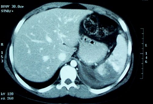
The image below shows likely areas of extravasation. They are a bit large to be pseudoaneurysms.

Spleen Blush-CT

Children are different than adults. Extravasation from spleen injuries in prepubescent children frequently stops on its own. Angiography should only be used if the child is failing nonoperative management.

Next post: A new paper looks at the natural history of these lesions.

VTE Prophylaxis Before Spine Surgery?

Many surgeons and surgical subspecialists are nervous about operating on people who are taking anticoagulants. This seems obvious when it involves patients on therapeutic anticoagulation. But it is much less clear when we are talking about lower prophylactic doses.

Spine surgeons are especially reluctant when they are operating around the spinal cord. Yet patients with spine injury are generally at the highest risk for developing venous thromboembolic (VTE) complications like deep venous thrombosis (DVT) or pulmonary embolism (PE). Is this concern warranted?

Surgeons at the Presley Trauma Center in Memphis examined this issue by performing a retrospective review of six years worth of patients who underwent spine stabilization surgery. They specifically looked at administration of any kind of preop prophylactic anticoagulant, and the most feared complications of bleeding complications and postop VTE.

Here are the factoids:

  • A total of 705 patients were reviewed, with roughly half receiving at least one preop prophylactic dose and the other half receiving none
  • There were 447 C-spine, 231 T-spine, and 132 L-spine operations, performed an average of 4 days after admission
  • Overall, bleeding complications occurred in 2.6% and VTE in 2.8%
  • Patients with VTE were more severely injured (ISS 27 vs 18)
  • Those who received at least half of their possible prophylactic doses had a significantly lower PE rate (0.4% vs 2.2%) but no significant difference in DVT or bleeding complications

Bottom line: So what to make of this? It’s a relatively small, retrospective study, and there is no power analysis. Furthermore, this hospital does not perform routine DVT screening, so that component of VTE may be underestimated, rendering the conclusions invalid.

However, the information on bleeding complications is more interesting, since this is much more reliably diagnosed using an eyeball check under the dressing. So maybe we (meaning our neurosurgeons and orthopedic spine surgeons) need to worry less about preop prophylactic VTE drugs. But we still need better research about whether any of this actually makes a dent in VTE and mortality from PE. To be continued.

Reference: Early chemoprophylaxis is associated with decreased venous thromboembolism risk without concomitant increase in intraspinal hematoma expansion after traumatic spinal cord injury. J Trauma 83(6):1108-1113, 2017.

A New Proposed Practice Guideline For Cervical Spine Clearance

In my last post, I reviewed a very recent prospective study on using CT scan alone for  cervical spine clearance in intoxicated patients. I believe that this is the final piece in the spine clearance puzzle to allow us to perform this task intelligently.

We’ve been accumulating more and more data that supports the use of CT scan in patients who fail clinical clearance. This failure can be due to the patient being obtunded or intoxicated, bearing a “distracting” injury, or being just plain uncooperative. Because of this, and our fear of missing a potentially devastating injury (typically because of rare anecdotal cases or urban legends), we have resorted to a significant degree of overkill. This has included, over the years, prolonged immobilization in a rigid collar, flexion/extension imaging (plain x-ray or fluoro), and MRI.

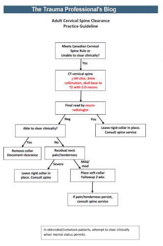
I’ve synthesized the available literature, and have drafted a simple, one sheet practice guideline for discussion. In order to use it, you must have the following:

  • A decent CT scanner – minimum 64 slice
  • A well-defined scan setup protocol – 3mm collimation, skull base to T2, 2-D reconstruction in sagittal and coronal planes (get a copy of our protocol below)
  • A skilled radiologist – neuroradiologist required

An image of the protocol can be found at the bottom of this post. I’m interested in your comments, and your comfort or discomfort with adopting something like this. Please leave comments here or on twitter.

Links: 

Reference: Cervical spine evaluation and clearance in the intoxicated patient: A prospective Western Trauma Association Multi-Institutional Trial and Survey. J Trauma 83(6):1032-1040, 2017.