Tag Archives: what the heck?

What The Heck? Pigtail Catheter Chest Tube

Here’s a case to make you think!

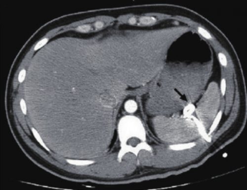
A patient arrives after being t-boned in his driver side door. He complains of left sided chest and abdominal pain. Chest x-ray shows a modest left hemopneumothorax. The decision is made to insert a pigtail type chest tube, and this is carried out in your trauma bay. It is uneventful, and a small amount of blood but no air is returned. The pelvis x-ray is unremarkable

The patient is then taken to CT, where an abdomen/pelvis scan with contrast is performed. This interesting slice is noted. What the heck?!

Here are my questions:

  • What is wrong in this picture?
  • How could it have been avoided?
  • Does a pigtail chest tube work for hemothorax?
  • How should this issue be managed, and where?

I’ll address these questions in my next post, and more!

Image source: internet

What The Heck? CT Imaging Problem: The Answer

I received some good guesses about this image yesterday, but no one got the right answer.

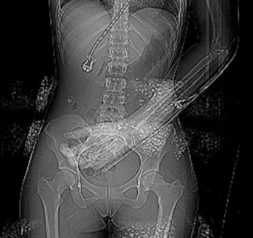
The patient had sustained blunt trauma and was undergoing CT imaging. The scout for the abdominal CT showed some kind of weird debris that interfered with the image, but when we uncovered and looked at the patient, nothing was visible:

What the heck? If you look carefully at the left side of the image, you can see that the “debris field” is on the surface of the patient. We can’t see in 3-D on images, but the difference in appearance on the left and right sides looks like it this stuff is wrapping around the patient.

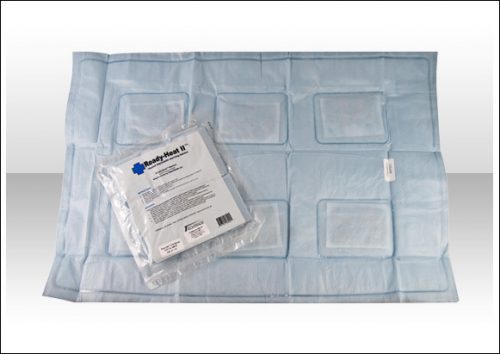
She was brought in by EMS with a warming blanket in place. On closer inspection, this was a thin, disposable blanket that heats up when removed from an airtight plastic pouch. These blankets contain thin pockets of a mineral mixture that looks like gravel. When exposed to air it heats up.

But on CT it looks like bone density material! When we looked at the patient, we were just lifting off the blanket that contained the offending material. Hence, we couldn’t find it.

Here’s a picture of one of these products. Note the six mineral pouches embedded in it., Don’t let this happen to you!


What The Heck? CT Imaging Problem

Here’s one for you. A patient is brought to you after a motor vehicle crash. You’ve completed your evaluation in the trauma resuscitation room, and you move off to CT for some imaging.

As the techs are preparing to do the abdominal CT, they perform the scout image to set up the study. This is what you see:

The arm was left down due to a fracture (note the splint along the forearm). But what is all that debris on the image? Other than a few abrasions here and there, nothing is visible on the skin in those areas.

What the heck? What do you think these are? Will they interfere with imaging? And what can you do about it?

Tweet or comment with your answers. I will explain all tomorrow.

What The Heck? Part 2: Progressive Back Pain After Heavy Lifting

Yesterday, I described a case of a young athlete who developed progressive back pain after rapidly increased his deadlift weights. He presented to the hospital with back pain and inability to get up from a supine position. He had firm and tender paraspinal muscles in his lower back, but no other findings.

What to do next? Obviously, we need a bit more information on the bony structures. Other than run of the mill muscle strain, a compression fracture would be the next most common diagnosis. In this young, healthy athlete, a simple set of AP and lateral spine images should be sufficient. But if you opted for a CT scan, I won’t argue. In either case, the images were normal.

Since there is significant muscle pain and tenderness, a lab panel with a few extras is in order, as well. The usual electrolytes, etc were normal. Creatinine was 0.9, but CPK was 60,000!

Now what are you thinking? What’s the diagnosis, and what is the decision tree for treatment?

Add your comments below, or tweet them out. I’ll finish this topic up in the next post.


What The Heck? Progressive Back Pain After Heavy Lifting

What the heck?! Here’s an interesting case of back pain! Can you figure it out?

A 20 year old male athlete has been performing 125 pound deadlift exercises recently. During his last session, he rapidly escalated to 6 reps at 235 pounds. He developed crampy lower back pain two hours later. The pain became rapidly worse, and he was evaluated at a hospital two days later.

He complained of unrelenting back pain, and could not get up or turn from a supine position. He denies taking any medications or supplements. There is no history of trauma.

On exam, he had firm and painful paraspinal muscles. Buttocks, thighs, and legs were nontender. All pulses were present. Straight leg raise and reverse straight leg raise tests were normal bilaterally. The abdomen was soft and nontender.

What are you thinking? What additional workup is needed at this point?

Post your comments below, or tweet them out. Tomorrow, we’ll walk through the diagnostic stuff, and Monday will be the big reveal.