Tag Archives: vascular

Best Of AAST 2021: Hard Signs Of Vascular injury

Well, it’s that time of year again! The annual American Association for the Surgery (AAST) is just a few weeks away. Starting today, I will begin reviewing some of the interesting abstracts (to me, at least) that will be presented. I’ll give my analysis and perspective, and usually provide some questions for the presenters that they may face during the live meeting. Enjoy!

I’ll start with abstract #1. This one is from the AAST Prospective Observational Vascular Injury Trial (PROOVIT).  The group was established to create an aggregate database of information on the presentation, diagnosis, management (acute and definitive), surveillance and outcomes following vascular trauma. It manages a registry that collects a wide variety of data on assorted vascular injuries.

This study re-examines our use of “hard signs” to diagnose vascular injury. Back in the day, we had “hard signs” and “soft signs.”  Hard signs were fairly obvious indicators of serious injury, such as pulselessness, ischemia, pulsatile bleeding, expanding hematoma, or a thrill or bruit. Soft signs were a bit less harsh: history of arterial bleeding, diminished pulse, stable hematoma, or an injury in proximity to the vessel.

In the old days, any hard sign of vascular injury was a hard indication to proceed directly to the OR for exploration and repair. However, the authors argue that in this day and age of advanced imaging and noninvasive treatment, maybe hard signs aren’t as hard as they used to be. They postulated that distinguishing between hemorrhage and ischemia would be more important in determining management of these injuries.

They focused on femoral and popliteal artery injuries, searching the database for classic hard vs soft signs, and newer ischemic (absent or diminished pulses, frank limb ischemia) vs hemorrhagic signs (overt hemorrhage, expanding hematoma, hypotension). They examined the presentation, pathology, treatment and outcome in 521 patients in the registry

Here are the factoids:

  • Hard signs occurred in 386, and 35% underwent CT angio instead of moving directly to OR
  • Soft signs occurred in the remaining 175, and 39% went to the OR without any further imaging
  • When using hemorrhage (HEM) vs ischemia (ISC), there were significant differences in mechanism (more penetrating in HEM), incidence of concomitant vein and nerve injury (higher in HEM), transection (higher in HEM), occlusion (higher in ISC)
  • For diagnosis and management, HEM was more likely to get intervention sooner, without imaging, using ligation or primary repair
  • ISC was more likely to undergo endovascular repair
  • HEM patients used a little more blood and had a higher mortality rate
  • Amputation rates, lengths of stay, and graft outcomes were the same

The authors concluded that the old hard vs soft signs paradigm no longer works, and suggest that using hemorrhage vs ischemia in now more useful.

Bottom line: This is a simple, straightforward descriptive study of five years of vascular injury of the proximal lower extremity. It certainly paints the picture that the old paradigm doesn’t work as well as it used to. About a third of patients with hard signs had preop imaging, and about the same number with soft signs went straight to OR.

The major drawback is that this is what I call a “how we do it study.” The results are largely dependent on the predominant practices at the participating centers. What if most of the centers that chose to participate are much more likely to use diagnostic imaging first, or go straight to OR first? And that centers that obeyed the classic hard vs soft signs paradigm steered clear? That could skew the results in this study.

This is a very thought-provoking paper. I’m looking forward to hearing more of the details at the meeting. I’ll be in the front row!

Questions for the authors and presenter:

  • Why did you focus only on femoral and popliteal injuries? What should be do about the others?
  • What were the “demographics” of the participating centers? What trauma center level, academic or not, urban or rural? All of these could have a significant impact on your numbers.
  • What was the duration of experience captured in the database? Are you able to see changes in preop eval or straight to OR practice over the years?

Reference: HARD, SOFT, & IRRELEVANT: HEMORRHAGIC & ISCHEMIC SIGNS BETTER DISTINGUISH IMPORTANT CHARACTERISTICS OF EXTREMITY VASCULAR INJURIES. AAST 2021, Oral abstract #1.


Best Of AAST #11: Hard Signs Of Vascular injury

The next abstract in this series poses a challenge to long-held dogma. More than three decades ago, examination of vascular injuries was divided into “hard signs” vs “soft signs.” Hard signs consisted of findings like pulsatile hemorrhage, expanding hematoma, absent distal pulses, thrill, or bruit. These were believed to be absolute indications to proceed directly to the operating room for exploration and repair.

But now, in this day and age of CT angiography  (CTA)and all manner of endovascular techniques and tools, things seem to be changing. There is more reliance on CTA, and a willingness to image patients with hard signs before considering an operation. But is this prudent?

The AAST established the Prospective Observational Vascular Injury Treatment (PROOVIT) database as a multi-institutional effort involving major trauma centers around the country in 2013. A group based at the Massachusetts General Hospital massaged the data to study current patterns in assessment and management of patients with penetrating extremity vascular injury. Specifically, they were interested in examining the presence of hard signs and the outcomes after initial imaging and operative management.

Here are the factoids:

  • A total of 1,910 database records were reviewed, of which 1108 (58%) presented with hard signs of injury
  • 83% of the patients with hard signs had either active hemorrhage or expanding hematoma; only 15% had ischemia
  • CTA was used in a quarter of patients with hard signs (24% hemorrhagic, 40% ischemic)
  •  Two thirds of patients with hard signs were taken to OR without imaging (70% hemorrhagic, 45% ischemic)
  • Open repair was performed in about two thirds of hemorrhagic and ischemic patients both with and without imaging, but endovascular  or hybrid repairs were 5x more likely (2% vs 10%) in patients who underwent imaging first
  • There were no differences in outcomes (amputation, mortality, blood transfusions, reoperation) between the open and endovascular/hybrid repair groups

The authors concluded that stable patients with hard signs of vascular injury may benefit from preop imaging to help plan the specific mode of repair to be performed (open vs endovascular / hybrid).

Here are my comments: This was a retrospective review of prospectively collected data. The database has a wealth of detail, and this is a simple and clean analysis of a specific question. The results and analyses were straightforward and easy to follow.

What this study does is to call into question the old dogma of rushing straight to the operating room with any patient who has hard signs of vascular injury. The advent of endovascular tools and techniques has allowed us to more easily address some vascular injuries that were previously problematic due to their location and accessibility.

Being a descriptive study only, it showed us “what we did” with vascular injuries during the time period of the database. And it also showed that the surgeons were more likely to use endovascular techniques if they were able to take the time for preop imaging. Most importantly, it demonstrated that gross outcomes like death, reoperation, and amputation were not increased by the delay needed to obtain that imaging.

I consider this to be a pilot project. And the authors correctly state that the next step is a true prospective study to confirm that this should be the new way of thinking about hard signs in the future.

Here are some questions for the presenter and authors.

  • Please provide more information on the database records used. Which years were included? What were the inclusion criteria? Were any patients excluded?
  • What was the definition of a vascular injury to the extremity? Did it include the very proximal brachial artery or the distal subclavian? These may increase the likelihood of choosing an endovascular repair.
  • Did you stratify by type of penetrating injury (stab vs gunshot) or velocity (assault rifles and shotguns)? These will increase the likelihood of proceeding directly to OR and potentially skew the data.
  • Some data from the abstract is missing, typically p values. This appears to be a glitch with the abstract entry system, since it is a problem in other abstracts as well.
  • How long do you think it will take to collect adequate data from a prospective study so that preop imaging in stable patients becomes the new standard of care?

This was a fun abstract to read! I’m looking forward to the presentation next week.

Vascular Trauma Resources At Pediatric Trauma Centers

There are two types of pediatric trauma centers: freestanding and combined. These adjectives refer to whether an adult trauma center is directly associated with the pediatric one. Over the years, I have come to appreciate that there may be substantial resource and experience differences between the two.

Trauma surgeons at freestanding centers are usually pediatric surgeons. They have managed trauma cases during their surgical residency and pediatric surgical fellowship, but usually have not taken a trauma fellowship. Their experience with complex trauma and advanced concepts like damage control surgery generally comes from their training and on the job experience. Surgeons at combined centers may be pediatric trained, or may be adult surgeons with pediatric experience. The adult surgeons are generally well-versed in advanced trauma concepts, and the pediatric surgeons can take advantage of the adult surgeons’ expertise in advanced trauma cases.

Freestanding pediatric centers may have fewer resources in some key areas, such as fellowship trained specialists in vascular surgery, GI endoscopy, and interventional radiology. A recent study accepted for publication from the University of Arkansas examines differences in surgeon practice patterns and resource availability at freestanding vs combined centers.

Two surveys were sent to 85 pediatric trauma centers around the US. Roughly half were Level I, and half were freestanding. One was sent to 414 pediatric surgeons at those centers inquiring about practice patterns, and the other was sent to the trauma medical directors of each center asking about their resources.

Here are the factoids:

  • 50 of the 85 trauma centers responded, as did 176 of the 414 surgeons. 48% of trauma medical directors responded. These are reasonable response rates for questionnaires.
  • Adult surgeons covered pediatric trauma at 6% of Level I centers, and 33% of Level II
  • During pediatric surgical fellowship, 56% participated in management of vascular trauma, 25% was managed by vascular surgeons, and 19% had no experience
  • At 23% of freestanding centers, vascular surgeons were not always available, and a vascular surgeon was not listed on the call schedule 38% of the time
  • 27% of freestanding facilities indicated that endovascular and stent capabilities were not available, and 18% did not have interventional radiologists (IR) available within 30 minutes
  • All combined centers had vascular and endovascular capabilities, and IR was available within 30 minutes 92% of the time

Bottom line: This is an intriguing paper that looks at a few of the disparities between freestanding and combined pediatric trauma centers. Obviously, it is hampered by the survey format, but does provide some interesting information. The focus was on vascular resources, and shows several of the major differences between the two types of centers.

Fortunately, vascular trauma is relatively rare in the pediatric age group. But it is possible that a child presenting to a freestanding pediatric trauma center may be managed by a pediatric surgeon with little vascular experience, and assistance from a fellowship trained vascular surgeon and/or interventional radiologist may be unavailable.

This paper provides important information regarding resource disparities in pediatric trauma care. Ideally, this should be reviewed and remedied as the Resources for Optimal Care of the Injured Patient (Orange Book) evolves over the coming years.

Reference: Pediatric Vascular Trauma Practice Patterns and Resource Availability: A Survey of ACS-Designated Pediatric Trauma Centers. J Trauma, accepted for publication Jan 12, 2018.

ABI vs API For Vascular Trauma

In general, the first maneuver in evaluating for possible vascular injury in an extremity is the good old physical exam. Is there a pulse or isn’t there? You can then subdivide that into: is the pulse weaker than normal. The problem is, what is “normal?” In most cases, we just compare it to another pulse somewhere and make a subjective judgment.

But we love to be more objective about things. Over the years, two simple, noninvasive techniques for evaluating pulses have been developed. The first is the ankle brachial index (ABI) , which was first described in 1930 and was used for diagnosis of peripheral vascular disease in 1950. It is performed by dividing the systolic pressure at the ankle of the affected extremity by the systolic pressure of one of the brachial arteries in the arms.

The new(er) kid on the block is the arterial pressure index (API), first described in 1991. This value is calculated by dividing the systolic pressure in the affected extremity by the systolic pressure in the contralateral uninjured extremity.

Many trauma professionals use the ABI when evaluating for potential vascular trauma. The typical threshold for pursuing further evaluation is 0.9, and several papers have been published on this topic. The API has also been critically evaluated, and the same threshold is used.

However, I believe that the API is more relevant and accurate than ABI. Why? Patients with atherosclerotic disease typically manifest it in their lower extremities. This serves to falsely elevate the ABI to a value greater than 1.0. It becomes more difficult to get down to that critical value of 0.9 that might indicate a vascular injury. Thus, the ABI may not detect a true injury, especially one in the lower extremities.

The API, on the other hand, relies on the fact that the amount of atherosclerotic disease is usually symmetric between the two lower extremities or the two upper extremities. Thus, the value will not be falsely elevated and will more accurately reflect the presence or absence of a vascular injury.

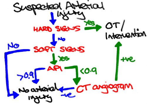
Bottom line: I recommend that you use the API when evaluating extremity vascular injury. Calculate the ratio by dividing the systolic pressure in the injured extremity with the pressure in the contralateral uninjured extremity (if there is one). A value < 0.9 indicates the need for angiographic evaluation, usually by CT scan.

And here’s a nice algorithm for managing peripheral vascular trauma from Life in the Fastlane:

Reference: Can Doppler Pressure Measurement Replace “Exclusion” Arteriography in the Diagnosis of Occult Extremity Arterial Trauma? Ann Surg 214(6):737-741, 1991.

Blunt Vertebral Artery Injury

Following up on yesterday’s post, I’ll deal with vertebral artery injuries today. These injuries are uncommon, making them hard to study and develop management recommendations. The literature suggests that about 1% of blunt trauma patients may sustain one of these. Most commonly, the method is motor vehicle crash, and just about any mechanism (hyperflexion, hyperextension, distraction injury, and facet fractures). Fracture of C1-3 has a higher association with the injury.

What is the natural history of this injury? If treated, 67% of occluded vessels recanalize, and 90% of stenotic arteries return to normal caliber. About 15% of untreated injuries will suffer a stroke. As seen in the paper cited yesterday, a good number of these are present on patient arrival and are nonpreventable. But the key issues are identifying an injury in the first place, and treating appropriately. Unfortunately, these are not straightforward.

Although the gold standard for detecting this lesion is digital subtraction angiography, no one does this in acute trauma patients anymore. CT angiography is well established, and the sensitivity rate approaches 99%. The main question is when to get it. To see my hospital’s interpretation of the literature, download our blunt imaging protocol below.

Treatment options include anticoagulation / antiplatelet therapy and endovascular therapy. There is much more experience with the former, but it can’t be used in patients at risk for bleeding (e.g. severe TBI). Unfractionated heparin is good for in-hospital use because it easily reversed. Longer term, anti-platelet agents are preferred. Aspirin is cheaper than clopidagrel, and no study has shown convincing superiority of one over the other. Determining whether endovascular stenting or embolization is necessary requires consultation with a neurosurgeon and interventional radiologist. The decision making is complex and not laid out in the literature. It’s flying by the seat of one’s pants, at best but can be a valuable adjunct.

Followup imaging is suggested to help determine when and if anti-platelet therapy can be discontinued. The best timing for these studies has not been worked out, but since these lesions tend to evolve over 7-10 days, any time after 2 weeks should be appropriate.

Bottom line: This is a tough topic because of the scarcity of good data, which in turn is due to the rarity of the injury. I believe that finding the lesions with good screening criteria offers the best chance of preventing complications such as stroke. Choice of management is best done in collaboration with your neurosurgical and radiologist colleagues.

Related posts: