Tag Archives: tips

Practical Tip: Penetrating Injury To The Vertebral Artery

This is an uncommon injury. But when encountered it can cause the trauma professional (and the patient) some major headaches. The majority of the vertebral artery injuries you are likely to encounter are caused by blunt trauma. They are generally diagnosed using CT angiography, and the treatment usually consists of low dose anti-platelet agents like aspirin. Occasionally, coiling or stenting using interventional radiology is needed.

But penetrating trauma is a totally different animal. Gunshot is the most common mechanism, because of the small windows available to access the artery within the vertebral canal using a knife. See the course of the artery in the picture below:

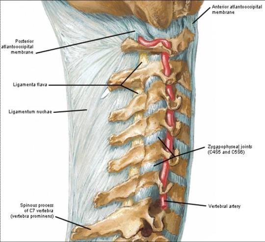
Unfortunately, this bony cage also makes it difficult to surgically approach the artery, especially if the field is continually filling with blood.

The techniques for dealing with this injury according to the doctor books are:

  • Send the patient to interventional radiology. Cutting off flow using coils is the preferred technique. Gelfoam and other products are not used because of the concern for distal embolization (to the brain). Stenting may be a consideration for blunt trauma, but not for penetrating.
  • Or, obtain proximal control by ligating the vertebral artery as it takes off from the subclavian. Hmm, this requires either a separate incision, or a supraclavicular extension of your neck incision. It takes time and is not as easy as it sounds.

Generally, the trauma surgeon stumbles upon this injury while doing a trauma neck exploration. Bleeding can be pesky, and may serve to obscure the field. My preferred method of control is:

  • Jam a wad of bone wax into the vertebral canal right where the bleeding is coming from.
  • Then jam another wad into the canal in the space below it. Proximal control!
  • Jam one final wad into the space above, if accessible. Distal control!

End of problem. Then do a thorough evaluation for all other injuries and address them. Feel free to share any additional tips that you may have!

Managing Penetrating Injuries: Some Practical Tips

Although penetrating injuries are a relatively uncommon mechanism at most trauma centers, they are more likely than not to injure deeper structures. Key decisions need to be made quickly during the initial evaluation in order to provide the best care.

Here are some practical tips:

  • Penetrating injuries to just about anything but the extremities should activate your trauma team.
  • If your patient is hypotensive, they will need to go to the OR. You can certainly start infusing some fluid or blood, but a lot leaked out before they got to you, indicating that the leak needs to be surgically fixed. No exceptions.
  • All hypotensive patients require activation of your massive transfusion protocol and consideration of giving tranexamic acid (TXA).
  • If your patient is normotensive, you have the luxury of evaluating them more thoroughly. But don’t lose your sense of urgency. Assume they are dying until you prove otherwise.
  • Complete your secondary survey. Don’t skimp on the exam and always look at the back. If your patient ends up on an OR table, it may be the only time you get to look at it for quite some time.
  • Get a single x-ray of the affected area, even if you need to go to the OR quickly. This can help plan your operation, and may drive you to explore areas you had not considered.
  • Before shooting the x-ray, mark any and all entry and exit points. This will help to predict the trajectory and any injured structures.
  • Use small markers, but not too small. Most radiology departments have small arrows, which are ideal. Dots are too small and may not show up well on plain images. But be aware that some markers may be too dense for CT, causing artifacts that may obscure pathology.
  • Watch out for your own safety! Somebody was trying to kill your patient, and they may show up at your hospital to try to finish the job. Make sure your ED and inpatient areas take appropriate security precautions.

The “Dang!” Factor

This tip is for all trauma professionals: prehospital, doctors, nurses, etc. Anyone who touches a trauma patient. You’ve probably seen this phenomenon in action. A patient sustains a very disfiguring injury. It could be a mangled extremity, a shotgun blast to the torso, or some really severe facial trauma. People cluster around the injured part and say “Dang! That looks really bad!”

It’s just human nature. We are drawn to extremes, and that goes for trauma care as well. And it doesn’t matter what your level of training or expertise, we are all susceptible to it. The problem is that we get so engrossed (!) in the disfiguring injury that we ignore the fact that the patient is turning blue. Or bleeding to death from a small puncture wound somewhere else. We forget to focus on the other life threatening things that may be going on.

How do we avoid this common pitfall? It takes a little forethought and mental preparation. Here’s what to do:

  • If you know in advance that one of these injuries is present, prepare your crew or team. Tell them what to expect so they can guard against this phenomenon.
  • Quickly assess to see if it is life threatening. If it bleeds or sucks, it needs immediate attention. Take care of it immediately.
  • If it’s not life threatening, cover it and focus on the usual priorities (a la ATLS, for example).
  • When it’s time to address the injury in the usual order of things, uncover, assess and treat.

Don’t get caught off guard! Just being aware of this common pitfall can save you and your patient!

But The Radiologist Made Me Do It!

The radiologist made me order that (unnecessary) test! I’ve heard this excuse many, many times. Do these phrases look familiar?

  1. … recommend clinical correlation
  2. … correlation with CT may be of value
  3. … recommend delayed CT imaging through the area
  4. … may represent thymus vs thoracic aortic injury (in a 2 year old who fell down stairs)
Some trauma professionals will read the radiology report and then immediately order more xrays. Others will critically look at the report, the patient’s clinical status and mechanism of injury, and then decide they are not necessary. I am firmly in the latter camp.
But why do some just follow the rad’s suggestions? I believe there are two major camps:
  • Those that are afraid of being sued if they don’t do everything suggested, because they’ve done everything and shouldn’t miss the diagnosis
  • Those that don’t completely understand what is known about trauma mechanisms and injury and think the radiologist does
Bottom line: The radiologist is your consultant. While they are good at reading images, they do not know the nuances of trauma. Plus, they didn’t get to see the patient so they don’t have the full context for their read. First, talk to the rad so they know what happened to the patient and what you are looking for. Then critically look at their read. If the mechanism doesn’t support the diagnosis, or they are requesting unusual or unneeded studies, don’t get them! Just document your rationale clearly in the record. This provides best patient care, and minimizes the potential complications (and radiation exposure) from unnecessary tests.
Related post: 

Reference: Pitfalls of the vague radiology report. AJR 174(6):1511-1518, 2000.

Tips For Surgeons: Seat Belt Sign

We see seat belt signs at our trauma center with some regularity. There are plenty of papers out there that detail the injuries that occur and the need for a low threshold for surgically exploring these patients. I have not been able to find specific management guidelines, and want to share some tidbits I have learned over the years. Yes, this is based on anecdotal experience, but it’s the best we have right now.

Tips for surgeons:

  • Common injuries involve the terminal ileum, proximal jejunum, and sigmoid colon. My observation is that location in the car is associated with the injury location, probably because of the location of the seat belt buckle. In the US, drivers buckle on the right, and I’ve seen more terminal ileum and buckethandle injuries in this group. Front seat passengers buckle on the left, and I tend to see proximal jejunum and sigmoid injuries more often in them.
  • Seat belt sign on physical exam requires abdominal CT for evaluation, regardless of age. The high incidence of significant injury mandates this test.
  • Seat belt sign plus any anomaly on CT requires evaluation in the OR. The only exception would be a patient with minimal fluid only in the pelvis with an unremarkable abdominal exam. But I would watch them like a hawk.
  • In patients who cannot be examined clinically (e.g. severe TBI), a rising WBC count or lactate beginning on day 2 after adequate resuscitation should prompt a trip to the OR. This is an indirect method for detecting injured bowel or mesentery.
  • Laparoscopy may be used in patients with equivocal findings. Excessive blood, bile tinged fluid, succus, or lots of fibrin deposits on the bowel should prompt conversion to laparotomy. Tip: place all ports distant to the seat belt mark. The soft tissues are frequently disrupted, and gas may leak into this pocket prohibiting good insufflation of the peritoneal cavity.
  • If in doubt, open the abdomen. It’s bad form to put in the scope, see something odd, and walk away. Remember, any abnormal finding after trauma is related to trauma until proven otherwise. It’s almost never pre-existing disease.

Related posts: