Tag Archives: Practice guideline

Best Practices For TBI Patients On Oral Anticoagulants: Part 1

Over the past five years, there has been a tremendous increase in the number of patients presenting to hospitals with traumatic brain injury. The bulk of these injuries occur in the elderly, and a rapidly growing number of them are taking anticoagulants for management of their medical comorbidities. Although there is a growing body of literature addressing this issue, many practical questions remained unanswered. This is due to the lack of randomized controlled studies of the clinical problems involved. And given the ethical issues of obtaining consent for them, there likely never will be.

An interdisciplinary group of Austrian experts was convened last year to consider the most common questions asked about TBI and concomitant anticoagulant use. They reviewed the existing literature from 2007 to 2018 and combined it with their own expertise to construct some initial answers to those questions.

Over the course of my next few posts, I’ll dig into each of the questions and review their suggested answers. And remember, all these Q&A apply to patients with known/suspected TBI with known/suspected oral anticoagulant use.

Let’s start with some diagnosis questions.

Q1. Should head CT be performed in all patients with known or suspected TBI and suspected or known use of anticoagulants?

Answer: All patients with TBI and potential or known use of anticoagulants should undergo an initial screening CT scan of the head.

A number of systems that predict the utility of head CT already exist (e.g. Canadian head CT rules). However, they do not and cannot take into account the various permutations of drugs and other medical conditions that may influence coagulation status. Vitamin K antagonists (VKA) like warfarin have been clearly shown to increase mortality after TBI. Data involving the use of anti-platelet agents or direct oral anticoagulants (DOAC) are a bit less clear.

Q2. Should a repeat head CT scan be repeated in these patients, and if so, when?

Answer: Patients with intracranial hemorrhage on their initial scan should have a repeat within 6-24 hours, based on the location of the bleed.

The natural course of patients who have an identified intracranial hemorrhage is extremely unpredictable. For that reason, a repeat scan is suggested. However, there are no consistent data that would indicate when this should occur. Indications and potential for progression vary by type of bleed (subarachnoid, subdural, epidural, intraparenchymal). Thus, you must work with your neurosurgeons to arrive at a reasonable repeat interval, and it may be different for a high-risk location (epidural) vs one with low risk (subarachnoid).

Q3. Should a patient with an initial head CT that is negative be admitted for neurologic monitoring?

Answer: Patients taking only aspirin with GCS 15 and initially negative head CT may be discharged. All other patients should be admitted for at least 24 hours for neurologic monitoring as follows (q1 hr x 4 hrs, q2 hr x 8 hrs, q4 hr x 12 hrs). Repeat head CT is indicated if there is any deterioration in neurologic exam.

Multiple papers have described the occurrence of delayed intracranial hemorrhage in patients taking oral anticoagulants other than aspirin. Although some bleeds may develop days or weeks after the initial injury, the majority occur during the first 24 hours. Routine repeat head CT in this group of patients with an initially negative scan has not been found to be helpful.

Q4. What about patients with an initially negative head CT who cannot be examined neurologically (intubation, sedation, dementia)?

Answer: Unexaminable patients should undergo a repeat head CT within 6-24 hours based on the underlying risk factors for development of delayed hemorrhage.

There is no real literature on this topic, but this statement makes sense. Each center should pick a reasonable time interval and include it in their own practice guideline.

In my next post, I’ll review the panel’s recommendations on coagulation tests and target levels for reversal of the various classes of anticoagulants.

Reference: Diagnostic and therapeutic approach in adult patients with traumatic brain injury receiving oral anticoagulant therapy: an Austrian interdisciplinary consensus statement. Crit Care 23:62, 2019.

Practice Guideline: Chest Tube Management (Part 2)

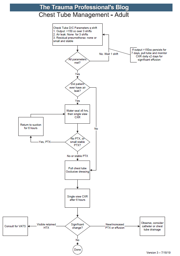
Yesterday, I went over the rationale for developing a practice guideline for something as simple and lowly as chest tube management. Today, I’m posting the details of the guideline that’t been in use at my hospital for the past 15 years. I’ve updated it to reflect two lessons learned from actually using it.

Here’s an image of the practice guideline. Click to open a full-size copy in a new window:

Here are some key points:

  • Note the decision tree format. This eliminates uncertainty so that the clinician can stick to the script. There are no hedge words like “consider” used. Just real verbs.
  • We found that hospital length of stay improved when we changed the three parameters from daily monitoring to three consecutive shifts. We are prepared to pull the tube on any shift, not just during the day time. And it also allows this part of the guideline to be nursing driven. They remind the surgeons that criteria are met so we can immediately remove the tube.
  • Water seal is only used if there was an air leak at some point. This allows us to detect a slow ongoing leak that may not be present during our brief inspection of the system on rounds.
  • The American College of Surgeons Committee on Trauma expects trauma centers to monitor compliance with at least some of their guidelines. This one makes it easy for a PI nurse or other personnel to do so.
  • The first of the “new” parts of this guideline is: putting a 7 day cap on failure due to tube output greater than 150cc per three shifts. At that point, the infectious risks of keeping a tube in begin to outweigh its efficacy. Typically, a small effusion may appear the day following removal, then resolves shortly.
  • The second “new” part is moving to VATS early if it is clear that there is visible hemothorax that is not being drained by the system. Some centers may want to try irrigation or lytics, but the data for this is not great. I’ll republish my posts on this over the next two days.

Click here to download a copy of this practice guideline for adults.

Click here to download the pediatric chest tube practice guideline.

Practice Guideline: Chest Tube Management (Part 1)

Management of chest tubes is one of those clinical situations that are just perfect for practice guideline development: commonly encountered, with lots of variability between trauma professionals. There are lots of potential areas for variation:

  • How long should the tube stay in?
  • What criteria should be used to determine when to pull it?
  • Water seal or no?
  • When should followup x-rays be done?

Every one of these questions will have a very real impact on that patient’s length of stay and potential for complications.

We developed a chest tube clinical practice guideline (CPG) at Regions Hospital way back in 2004! Of course, there was little literature available to guide us in answering the questions listed above. So we had to use the clinical experience and judgment of the trauma faculty to settle on a protocol that all were comfortable with.

Ultimately, we answered the questions like this:

  • The tube stays in until three specific criteria are met
  • The criteria are: <150 cc drainage over 3 shifts, no air leak, and no residual pneumothorax (or at least a small, stable one)
  • Use of water seal is predicated on whether there was ever an air leak
  • An x-ray is obtained to determine whether any significant pneumo- or hemothorax is present prior to pulling the tube, and 6 hours after pulling it

This CPG has been in effect for over 15 years with excellent results and dramatically shortened lengths of stay.  However, as with any good practice guideline, it needs occasional updates to stay abreast of new research literature or clinical experiences. We recognized that occasional patients had excessive drainage for an extended period of time. This led us to limit the length of time the tube was in to seven days. And we also noted that a few patients had visible hemothorax on their pre-pull imaging. These patients were very likely to return with clinical symptoms of lung entrapment, so we added a decision point to consider VATS at the end of the protocol.

I’ll share the full protocol tomorrow and provide a downloadable copy that you can modify for your own center. I’ll also give a little more commentary on the rationale for the key decision points in this CPG.

Related posts:

Practice Guidelines And Tincture Of Time

Most trauma centers have at least a few practice guidelines to help the standardize the way they manage common injuries. Solid organ injury. Elder trauma. Chest tube management. But they are all designed for use in patients who present shortly after their injury.

What about someone who presents a day or two, or more, after their injury?  That changes the picture entirely. Most guidelines have a time component built in. A TBI protocol requires a repeat head CT after a certain period of time. Solid organ injury patients may have restricted activity or frequent vital signs for a while.

But all too often, trauma professionals treat the patient with delayed presentation exactly the same as fresh trauma. For example, a patient falls and bumps their head. They have a persistent headache, and after two days decide to visit their local ED. The CT scan shows a small amount of subarachnoid blood in the area of the impact. Your practice guidelines says to admit for observation, frequent neruo checks, and repeat head CT in 12 hours.

Or a young male playing sports took a hit to his left flank. After 3 days, he’s just tired of the pain and comes to the ED for some pain medication. CT scan shows a grade III spleen injury with a small amount of hemoperitoneum. Your protocol says to admit, make NPO, liimit activity, and observe for 2 days.

What would I do in these cases? Think about it! If the patients had presented right after the event, they would have gone through your guideline and would have been discharged already. So I would review the images, talk to the patients about their injuries, then send them home from the ED with followup. They’ve already passed!

Bottom line: Remember, practice guidelines are not etched in stone. Variances are possible, but need to be well thought out in advance. And hopefully documented in the chart to expedite the inevitable trauma performance improvement inquiry. If the requisite amount of time has gone by, and the history and exam are reasonable, the patient has already passed your protocol. Send them home.

Related posts:

How To Craft A Clinical Practice Guideline

All US trauma centers verified by the American College of Surgeons are required to have clinical practice guidelines (CPG). Trauma centers around the world generally have them, but may not be required to by their designating authority. But don’t confuse a policy about clinical management, say for head injury, with a real CPG. Policies are generally broad statements about how you (are supposed to) do things, whereas a CPG is a specific set of rules you use when managing a specific patient problem.

  1. Look around; don’t reinvent the wheel! This is the first mistake nearly every center makes. It seems like most want to spend hours and hours combing through the literature, trying to synthesize it and come up with a CPG from scratch. Guess what? Hundreds of other centers have already done this! And many have posted theirs online for all to see and learn from. Take advantage of their generosity. Look at several. Find the one that comes closest to meeting your needs. Then “borrow” it.
  2. Review the newest literature. Any existing CPG should have been created using the most up to date literature at the time. But that could have been several years ago. Look for anything new (and significant) that may require a few tweaks to the existing CPG.
  3. Create your draft, customizing it to your hospital. Doing things exactly the same as another center doesn’t always make sense, and it may not be possible. Tweak the protocols to match your resources and local standards of care. But don’t stray too far off of what the literature tells you is right.
  4. Make sure it is actionable. It should not be a literature summary, or a bunch of wishy-washy statements saying you could do this or consider doing that. Your CPG should spell out exactly what to do and when. (see examples below)
  5. Create a concise flow diagram. The fewer boxes the better. This needs to be easy to follow and simple to understand. It must fit on one page!
  6. Get buy-in from all services involved. Don’t try to implement your CPG by fiat. Use your draft as a launching pad. Let everyone who will be involved with it have their say, and be prepared to make some minor modifications to get buy-in from as many people as possible.
  7. Educate everybody! Start a campaign to explain the rationale and details of your CPG to everyone: physicians, nurses, techs, etc. Give educational presentations. You don’t want the eventual implementation to surprise anyone. Your colleagues don’t like surprises and will be less likely to follow along.
  8. Roll it out. Create processes and a timeline to roll it out. Give everyone several months to get used to it.
  9. Now monitor it! It makes no sense to implement something that no one follows. Create a monitoring system using your PI program. Include it in your reports or dashboards so providers can see how they are doing. And if you really want participation, let providers see how they are doing compared to their colleagues. Everyone wants to be the top dog.

Some sample CPGs: