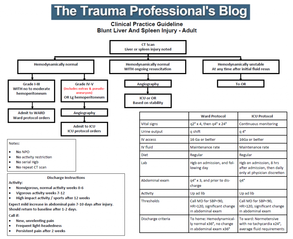
Tag Archives: liver

Post-Embolization Syndrome In Trauma

A reader requested that I write about post-embolization syndrome. Not being an oncologist or oncologic surgeon, I honestly had never heard about this before, let alone in trauma care. So I figured I would read up and share. And fortunately it was easy; there’s all of one paper about it in the trauma literature.

Post-embolization syndrome is a constellation of symptoms including pain, fever, nausea, and ileus that occurs after angio-embolization of the liver or spleen. There are reports that it is a common occurrence (60-80%) in patients being treated for cancer, and there are a few papers describing it in patients with splenic aneurysm. But only one for trauma.

Children’s Hospital of Boston / Harvard Medical School retrospectively reviewed 12 years of their pediatric  trauma registry data. For every child with a spleen injury who underwent angio-embolization, they matched four others with the same grade of injury who did not. A total of 448 children with blunt splenic injury were identified, and (thankfully) only 11 underwent angio-embolization. Nine had ongoing bleeding despite resuscitation, and two had developed splenic pseudoaneursyms.

Here are the factoids:

  • More of the children who underwent embolization had extravasation seen initially and required more blood products.  They also had longer ICU (3 vs 1 day) and hospital stays (8 vs 5 days). Not surprising, as that is why they had the procedure.
  • 90% of embolized kids had an ileus vs 2% of those not embolized, and they took longer to resume regular diet (5 vs 2 days)
  • Respiratory rate and blood pressure were higher on days 3 and 4 in the embolized group, as was the temperature on day 5 (? see below)
  • Pain was higher on day 5 in the embolized group (? see below again)

Bottom line: Sorry, but I’m not convinced. Yes, I have observed increased pain and temperature elevations in patients who have been embolized. Some have also had an ileus, but it’s difficult to say if that’s from the procedure or other injuries. And this very small series just doesn’t have enough power to convince me of any clinically significant differences in injured children.

Look at the results above. “Significant” differences were only identified on a few select days, but not on the same days across charts. And although the authors may have demonstrated statistical differences, are they clinically relevant? Is a respiratory rate of 22 different from 18? A temp of 37.8 vs 37.2? I don’t think so. And length of stay does not reveal anything because the time in the ICU or hospital is completely dependent on the whims of the surgeon.

I agree that post-embolization syndrome exists in cancer patients. But the findings in trauma patients are too nondescript. They just don’t stand out well enough on their own for me to consider them a real syndrome. As a trauma professional, be aware that your patient probably will experience more pain over the affected organ for a few days, and they will be slow to resume their diet. But other than supportive care and patience, nothing special need be done.

Related posts:

Reference: Transarterial embolization in children with blunt splenic injury
results in postembolization syndrome: A matched
case-control study. J Trauma 73(6):1558-1563, 2012.

Does Trauma Center Level Make A Difference In Treating Solid Organ Injury?

In the last two posts, I reviewed contrast anomalies in solid organs, specifically the spleen. Today, I’ll be more general and examine a recent paper that compared management and outcomes after the other major solid organ injury, liver, at Level I vs Level II trauma centers.

There are several papers that have detailed overall differences in outcomes, and specifically mortality, at Level I and II centers. Some of these show outcomes that are not quite as good at Level II centers when compared to Level I. On paper, it looks like these two levels should be very similar. Take away research and residents, and maybe a few of the more esoteric capabilities like reimplantation, and aren’t they about the same?

Well, not really. They can be, though. Level I criteria are fairly strict, and the variability between difference Level I centers is not very great. Level II criteria are a bit looser, and this allows more variability. Many Level II centers function very much like a Level I, but a few are only a bit higher functioning than a Level III with a few extra surgical specialists added in.

A paper currently in press used the Michigan Trauma Quality Improvement Program (MTQIP) data from all 29 ACS verified Level I and II centers in the state (wow!). Six years of information was collected, including the usual demographics, outcome data, and management. A total of 538 patients met inclusion criteria, and this was narrowed down to 454 so statistical comparisons of similar patients could be made for Level I vs Level II centers.

Here are the factoids:

  • Mortality was significantly higher in Level II centers compared to Level I (15% vs 9%) and patients were more likely to die in the first two days, suggesting hemorrhage as the cause
  • Patients were more likely to die in the ED at Level II centers, despite a significantly lower Injury Severity Score (ISS)
  • Pneumonia and ARDS were significantly more likely to develop in Level II center patients
  • Level II centers used angiography less often and took patients to the OR more frequently
  • Level II centers admitted fewer patients to the ICU, but ICU admission was associated with significantly decreased mortality
  • Complications were fewer at Level II centers, but they were less likely to rescue patients when they occurred

Bottom line: Level I and II centers are supposed to be roughly the same, at least on paper. But a number of studies have suggested that there are more disparities than we think. Although this paper is a retrospective review, the sheer number of significant differences and its focus on one particular injury makes it more compelling.

So what to do? Tighten up the ACS Orange Book criteria? That’s a slow and deliberate process that won’t help our patients now. The quickest and most effective solution is for all centers to adopt uniform practice guidelines so they all perform like the highly successful Level I programs in the study. There are plenty of them around. If you are not yet using one, I urge you to have a look at the example below. Tweak it to fit your center. And use your PI program to trend the outcomes!

Related post:

Reference: Variability in Management of Blunt Liver Trauma and Contribution of Level of ACS-COT Verification Status on Mortality. J Trauma, in press, Dec 1, 2017.

Trauma Residents: How To Remember Liver Anatomy

In trauma surgery, operative management of liver injury is usually messy business, with little time for nice anatomic resections. However, an understanding of the basic anatomy, especially that of the vascular supply is crucial for saving your patient.

A cool tool for remembering Couinaud’s segments and the overall layout of liver anatomy was published in the Archives of Surgery recently. It makes use of a model, which consists of your hand! Just make a fist with your right hand and tuck the thumb behind the other fingers.


The fingers can then be numbered according to the Couinaud segments, with the caudate lobe (segment 1) represented by the thumb that is tucked away. The PIP joints represent the plane that the portal vein runs through, with branches going to upper and lower segments. Note how the ring finger normally lies a little more anterior than the little finger in this position, just like the sectors of the right lobe.

The creases between the fingers represent the left, middle and right hepatic veins.


The right hepatic vein is located between the right anterior and posterior sectors and the left hepatic vein sits between the left medial and lateral sectors. The middle hepatic vein is in between the left and right hemi-liver.

Bottom line: This “handy” liver model is available immediately in the OR and is already sterile. It can help visualize liver structures that may be injured quickly and accurately to speed your operative approach to the problem.

Reference: A Handy Tool to Teach Segmental Liver Anatomy to Surgical Trainees. Arch Surg 147(8):692-693, 2012.

Post-Embolization Syndrome In Trauma

A reader requested that I write about post-embolization syndrome. Not being an oncologist or oncologic surgeon, I honestly had never heard about this before, let alone in trauma care. So I figured I would read up and share. And fortunately it was easy; there’s all of one paper about it in the trauma literature.

Post-embolization syndrome is a constellation of symptoms including pain, fever, nausea, and ileus that occurs after angio-embolization of the liver or spleen. There are reports that it is a common occurrence (60-80%) in patients being treated for cancer, and there are a few papers describing it in patients with splenic aneurysm. But only one for trauma.

Children’s Hospital of Boston / Harvard Medical School retrospectively reviewed 12 years of their pediatric  trauma registry data. For every child with a spleen injury who underwent angio-embolization, they matched four others with the same grade of injury who did not. A total of 448 children with blunt splenic injury were identified, and (thankfully) only 11 underwent angio-embolization. Nine had ongoing bleeding despite resuscitation, and two had developed splenic pseudoaneursyms.

Here are the factoids:

  • More of the children who underwent embolization had extravasation seen initially and required more blood products.  They also had longer ICU (3 vs 1 day) and hospital stays (8 vs 5 days). Not surprising, as that is why they had the procedure.
  • 90% of embolized kids had an ileus vs 2% of those not embolized, and they took longer to resume regular diet (5 vs 2 days)
  • Respiratory rate and blood pressure were higher on days 3 and 4 in the embolized group, as was the temperature on day 5 (? see below)
  • Pain was higher on day 5 in the embolized group (? see below again)

Bottom line: Sorry, but I’m not convinced. Yes, I have observed increased pain and temperature elevations in patients who have been embolized. Some have also had an ileus, but it’s difficult to say if that’s from the procedure or other injuries. And this very small series just doesn’t have enough power to convince me of any clinically significant differences in injured children.

Look at the results above. “Significant” differences were only identified on a few select days, but not on the same days across charts. And although the authors may have demonstrated statistical differences, are they clinically relevant? Is a respiratory rate of 22 different from 18? A temp of 37.8 vs 37.2? I don’t think so. And length of stay does not reveal anything because the time in the ICU or hospital is completely dependent on the whims of the surgeon.

I agree that post-embolization syndrome exists in cancer patients. But the findings in trauma patients are too nondescript. They just don’t stand out well enough on their own for me to consider them a real syndrome. As a trauma professional, be aware that your patient probably will experience more pain over the affected organ for a few days, and they will be slow to resume their diet. But other than supportive care and patience, nothing special need be done.

Related posts:

Reference: Transarterial embolization in children with blunt splenic injury
results in postembolization syndrome: A matched
case-control study. J Trauma 73(6):1558-1563, 2012.

Incidental Finding: Gas In The Spleen After Embolization

Most solid organ injury practice guidelines include angioembolization in part of the pathway. But very few require re-imaging at any point to see how the liver or spleen are coming along.

But every once in a while another condition arises, or symptoms worsen unexpectedly, causing us to get another CT scan that includes the abdomen and pelvis. And sometimes we see things that we wouldn’t normally see, like air bubbles in the organ that was embolized.

So what is okay, and what requires some kind of intervention? Our friends at ShockTrauma in Baltimore looked at this in 2001 and can provide some pretty good guidance. They reviewed patients who underwent CT scan both before and after embolization over about 2.5 years. They performed the post-embolization scans for specific indications like fevers, elevated WBC count (!), increasing abdominal pain, or an episode of hypotension. A total of 53 patients were studied.

Here are the factoids:

  • 24 patients underwent embolization of the main splenic artery, 22 had selective embolization of part of the spleen, and 7 had both
  • Splenic infarcts occurred in 63% of patients with main artery embolization, but were large (> 50% of the parenchyma) in only 20% of those
  • Infarcts occurred in 100% of selective embolizations, but were small (< 50%) in 93% of cases
  • Infarcts occurred in 71% of patients with both main and selective embolization, and most were small (80%)
  • Seven (13%) patients developed gas bubbles in the spleen, and was usually present for 1-7 days before disappearing
  • One patient developed increasing gas with pneumoperitoneum and underwent splenectomy for a splenectomy for abscess

This picture that shows tiny bubbles in the spleen parenchyma that represent “normal” gas after embolization:

And the following one shows an air/fluid collection in the spleen that indicates an abscess:

Bottom line: Tiny bubbles in the spleen (and probably the liver) occur normally after angioembolization. They usually develop within an area of infarction, and most are benign. It is possible for them to evolve into a splenic abscess, but unlikely. Many embolization patients develop fevers at some point, and most have an elevated WBC count. So in most cases, you can ignore this incidental finding, as long as your patient has mild symptoms.

However, if the patient develops high fevers, very elevated WBC (> 25K), increasing abdominal or flank pain, and the spleen develops an air/fluid level, an abscess is forming. Despite what your radiologist might suggest, catheter drainage is not a good idea. The tubes are too small to remove the slurry that is generally found within the abscess. A trip to the OR is the only effective treatment, and splenectomy is generally the only option.

Related posts:

Reference: CT Findings after Embolization for Blunt Splenic Trauma. J Vasc Intervent Radiol 12(2):209-214, 2001.