Tag Archives: DVT

Unfractionated vs Low Molecular Weight Heparin For Trauma Patients

In my last post, I described some of the telltale signs that could be seen in a trauma center’s TQIP report that might suggest there are issues with how they go about providing prophylaxis for venous thromboembolism for their patients. Today, I will analyze a systematic review and meta-analysis of a collection of research that compared the efficacy and safety of unfractionated heparin (UFH) to low molecular weight heparin (LMWH) specifically for trauma patients.

First, it’s important to understand the concept of research quality. There is a huge amount of research published these days, and it varies considerably in how well it is designed, executed, and analyzed. Here is a diagram that illustrates the levels of quality and the volume of research published at each level. By quality, I mean the applicability to clinical treatment of actual humans. For this reason, test tube and animal research are low on the pyramid.
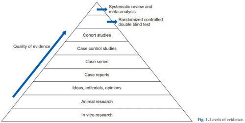
The research that most people consider to be the “gold standard” (randomized, controlled, double blind) is very close to the top. There is one class that, if conducted properly, may even be better. That is the systematic review and meta-analysis.

Most people have heard of meta-analysis, and it can be very good by itself. This combines lots of smaller studies into one larger one. However, it may hampered by the quality of the studies included in the meta-analysis. The tenet of “garbage in equals garbage out” certainly holds. But a systematic review takes that one step further.

The systematic review compiles all possible studies related to a small set of research questions, and usually concentrates on the ones with the highest quality research design. The quality of each of the studies is evaluated, and a meta-analysis is then performed on the best. Results are usually represented in a forest plot. This is an easy way to illustrate the estimated results from a number of studies that address the same question. There is also an entry that shows the relative strength of all of the studies combined. Here’s an example:

There are seven studies included, and each is displayed with its risk ratio (RR) and confidence interval (CI). The final diamond is the combined RR and CI for the entire group of studies. In the example above, note that most of the studies have CI bars that extend over the risk ratio = 1 line, meaning they may not be significant. But when taken together, the final risk ratio of the group is well under 1.0 and does not cross over it, denoting significance.

Let’s now apply this concept to a group of studies comparing UFH and LMWH for prevention of VTE for trauma patients. Based on keyword search, the authors identified 1,227 records for screening. Of those, only 40 were tentativley found to directly address the question. After in-depth analysis, only 12 were eligible for final review. For various reasons, only about 1 in 100 papers could be used to try to analyze the question. This always shocks me.

Here are the efficacy results. All are statistically significant, and all but mortality were stated with moderate certainty. The mortality number had low certainty due to the fact there were only three studies and confidence intervals were very wide.

  • Deep venous thrombosis: LMWH reduced by about 35% compared to UH
  • Pulmonary embolism: LMWH reduced by 44% although certainty was low
  • Any VTE: LMWH reduced by about 30%
  • Mortality: LMWH reduced by 56% (low to very low certainty)

Safety was also analyzed, including bleeding events, unexpected return to OR, heparin induced thrombocytopenia (HIT), and “any adverse events.” All of the Total Confidence Interval diamonds were situated on the risk ratio = 1 line, denoting no significant change when comparing LMWH vs UH.  However, quality of this data was noted to be low due to the quality of the individual studies. This means that we do not really know the answer to the safety question with any certainty yet.

Bottom line: This is one of the best summaries of our research on UH vs LMHW to date. It broadly reviewed the available literature and found only a small subset to analyze. It is clear that LMWH is superior for prevention of DVT and VTE overall. However, the impact on pulmonary embolism and death is still unclear.

As far as safety, the studies are still of quality that is too low to use for a decent analysis. Although this study did not detect any increase in complications, we still can’t say with any degree of certainty.

So what does it all mean? We have been using LMWH for decades now. Most likely, if there were regular complications like bleeding, unexpected return to OR, or HIT we would have definitely noticed it by now. Fortunately, we only have a few anecdotes and case reports to scare us off.

Overall, there is good support for the use of LMWH exclusively in most trauma patients. However, the prescribing provider should always assess patient factors that may suggest that UH might be better is a specific case. But remember that using UH trades an unclear/unlikely safety advantage for a recognized decrease in efficacy.

Reference: Efficacy and safety of low molecular weight heparin versus unfractionated heparin for prevention of venous thromboembolism in trauma patients. Ann Surgery 275(1):19-28, 2022.

Best Of EAST #6: How Long Does Risk For VTE Last After Spine Fracture?

Most trauma centers use an existing venous thromboembolism (VTE) guideline or have developed their own injury-specific one. These include risk factors, contraindications, specific agent, and dosing recommendations. But one thing most do not include is duration of prophylaxis!

The length of time a patient is at risk for VTE is not well delineated yet. The group at the University of Arizona decided to tackle this program using the National Readmission Database. This dataset is a comprehensive resource for critically analyzing patients who are discharged and readmitted, even for multiple occurrences. It covers 30 states and almost two thirds of the population.

The authors focused on VTE occurring during the first six months after injury. Patients who died on the initial admission, were taking anticoagulants, had spinal surgery, or sustained a spinal cord injury were excluded. Over 41,000 records from the year 2017 met these criteria.

Here are the factoids:

  • The average age was 61, which shows the skew toward the elderly with these injuries
  • Spine areas injured were cervical in 20%, thoracic in 19%, lumbar in 29%, sacrococcygeal in 11%, and multiple levels in 21%.
  • During the initial admission, 1.5% developed VTE: 0.9% were DVT and 0.7% were PE
  • Within 1 month of discharge, 0.6% of patients were readmitted for VTE: 0.4% DVT and 0.3% PE
  • In the first 6 months, 1.2% had been readmitted: 0.9% DVT and 0.6% PE
  • Mortality in the first 6 months was 6.7%
  • Factors associated with readmission for VTE included older age, discharge to a skilled nursing facility, rehab center, or care facility

The authors concluded that VTE risk remains high up to 6 months after conservatively managed spinal fractures. They recommend further study to determine the ideal prophylactic agent and duration.

Bottom line: This is a creative way of examining a difficult problem. We know that VTE risk does not stop when our patient is discharged. This is one of the few ways to get a sense of readmissions, even if it is not to the same hospital. And remember, this is an underestimate because it’s possible for a patient living near a state border to be re-hospitalized in a state not in this database.

This study might prompt us to prescribe up to six months of prophylaxis, particularly in seniors who are discharged to other care facilities.

Here are my questions for the author and presenter:

  • Is there any way to extrapolate your data to the entire population of the US, or to compensate for the “readmission over state lines” problem?
  • Is the odds ratio of 1.01 for risk of VTE in the elderly age group significant in any way? It seems like a very low number that would be easily overwhelmed by the “noise” in this data set.
  • Is the mortality number for all causes, or just VTE?

This is an intriguing study, and one that should influence the VTE guidelines in place at many trauma centers!

Reference: THE LONG-TERM RISKS OF VENOUS THROMBOEMBOLISM AFTER NON-OPERATIVELY MANAGED SPINAL FRACTURE. EAST 35th ASA, oral abstract #28.

Routine Duplex Screening For Venous Thromboembolism

Venous thromboembolism (VTE) is a potential problem for all hospitalized patients, and traumatic injury is yet an additional risk factor for its occurrence. Most trauma centers have some kind of risk assessment tool to help the tailor their chemoprophylaxis regimen to patients most at risk. But far fewer have adopted the use of screening ultrasounds to monitor for new onset VTE that would dictate conversion to therapeutic treatment.

Unfortunately, in the US, the Centers for Medicare and Medicaid Services (CMS) has deemed VTE as a “never” event and penalizes hospitals when they report it. One of the unintentional consequences of this (or is it?) is that hospitals may then pressure trauma programs to avoid surveillance in order to “make the numbers look better.” Remember Law X from Samuel Shem’s House of God?

X. If you don’t take a temperature, you can’t find a fever.

Similarly, if you don’t do a duplex screen, you probably won’t detect VTE. Now granted. some patients develop classic symptoms like edema, pain, and tenderness. But not that many.

But is this wise? My contention has been that if the patient doesn’t develop symptoms that catch your attention, yet they develop VTE that you don’t know about, they are at risk for more serious complications like pulmonary embolism (PE). And you are blithely unaware.

The trauma group at Intermountain Medical Center in Salt Lake City performed an elegant study to determine the impact of screening for VTE in their trauma patients. They performed a prospective, randomized trial on trauma patients admitted over a 30-month period. Patients were included if they were judged to be at moderate to high risk based on their risk assessment profile (RAP) score. Patients were excluded if they were children, had VTE or PE within 6 months prior to hospitalization, or had some type of hypercoagulable state.

Patients were sequentially randomized to no duplex screening vs screening on days 1, 3, 7, and then weekly thereafter. The primary outcome measure was PE during the hospital stay. Secondary outcomes consisted of a number of factors relating to development of DVT.

Here are the factoids:

  • Nearly two thousand patients were enrolled, with about 995 patients in each group and no differences in demographics
  • The ultrasound group had significantly more below-knee (124 vs 8) and above knee (19 vs 8) DVT identified (no surprise there)
  • The ultrasound group had significantly fewer pulmonary emboli than the no ultrasound group (1 vs 9) (lots of surprise here!)
  • Mortality was similar during the hospital stay and for 90 days after

Bottom line: If you look for it, you will find it! This is the definition of surveillance bias. But in in this study, looking for clots in the legs may also decrease the number of patients who develop symptomatic pulmonary embolism. How could this be?

There are a few possibilities. The majority of DVT found in the surveillance group were located distally. Although there is some uncertainty as to how likely these are to embolize, it is probably very low. So let’s ignore them for now and assume that only the proximal clots might embolize.

This leaves an extra 11 DVT found in the surveillance group over and above the no-ultrasound group. Despite that, the surveillance group had only one PE vs 9 in the no-ultrasound group!

Another explanation was that the ultrasound guided changes in management, shifting to management to therapeutic drug dosing. The authors did not find a significant difference between the use of therapeutic vs prophylactic dosing between the groups. But there was a difference. Although the overall study was well-powered, there really weren’t enough numbers to show whether there was a true difference in therapeutic dosing. Fourteen patients in the ultrasound group got therapeutic anticoagulation compared to only 4 in the no-surveillance group. I think this is the actual reason.

Overall, this is a well-designed and well-executed study that shows why taking the Ron Popeil approach to DVT prophylaxis (“set it and forget it”) doesn’t work. Patients do occasionally develop proximal DVT on standard chemoprophylaxis (and frequently develop distal DVT), but it doesn’t always result in obvious signs and symptoms. This study shows that if you don’t look for it, you may not know until they suddenly develop chest pain, air hunger, and worse! So consider carefully if your practice guideline doesn’t yet include surveillance.

Reference: Trauma Patients at Risk for Venous Thromboembolism who Undergo Routine Duplex Ultrasound Screening Experience Fewer Pulmonary Emboli: A Prospective Randomized Trial. J Trauma, publish ahead of print, Publish Ahead of Print. DOI: 10.1097/TA.0000000000003104, February 4, 2021.

Should I Apply Compression Devices To Patients With DVT?

Everyone knows that venous thromboembolism (VTE) is a potential problem in hospitalized patients, and especially so in trauma patients. Several groups of them are at higher risk by virtue of the particular injuries they have sustained and the activity restriction caused.

Nearly every trauma program uses some form of screening and prophylaxis in an attempt to reduce the occurrence of this problem, which can result in deep venous thrombosis (DVT) and/or pulmonary embolism (PE). Screening looks at patient factors such as age, obesity, previous VTE as well as injury risk factors like spine and pelvic fractures, and decreased mobility.

Based on the screening protocol, prophylaxis may be prescribed depending upon level of VTE risk, which is then balanced with bleeding risk from brain, solid organ, or other injuries. The choices we have are primarily mechanical vs chemical and consist of compression devices (sequential or not) and various heparins.

An age old question surfaced on my own patient rounds recently. If a patient breaks through their prophylaxis and develops DVT, is it safe to apply compression devices to the extremity?

There has always been the fear that doing things that increase flow in the affected extremity may cause clots to dislodge and ultimately cause a PE. Seems logical right? But we know that often, our common sense about things is completely wrong.  Couldn’t just moving around cause pieces to break off? A meta-analysis of 13 studies published in 2015 showed that early ambulation was not associated with a higher incidence of new PE. Furthermore, patients who suffered from pain in the affected extremity noted significant improvements with early ambulation.

If ambulation makes the pain better, could the veins be recanalizing more quickly? Another study examined a small group of 72 people with DVT receiving anticoagulants, half of whom were prescribed exercise and compression stockings and the other half stockings only. There was a huge amount of variability in the rates of recanalization, but ultimately there were no significant differences with or without exercise.

So just lying in bed is not good, and exercise/ambulation may actually make people feel better. But interestingly, bedrest alone does not appear to increase the likelihood of PE! It does decrease the risk of developing problems other than the VTE, like pulmonary complications.

But what about compression devices? Common sense would say that you are intermittently  increasing pressures in the leg veins, which could dislodge any loose clots and send them flying to the lungs, right?

Unfortunately, I couldn’t find a paper from anyone who had the courage to try this. Or perhaps no institutional review board (IRB) would approve it. But the key fact is that every compression device manufacturer includes existing DVT as a contraindication in their product documentation. They don’t have any literature either, so I assume it’s an attempt to limit litigation, just in case.

Bottom line: Walking provides at least as much muscle compression as compression devices. But the simple truth is that we have no solid research that either supports or condemns the use of active compression devices in patients with known DVT. And we probably won’t, ever.

Compression stockings seem to be safe, but they really don’t do much. They are white, but don’t do much more than contribute to hospital clothing fashion. Since the manufacturers define existing DVT as a contraindication, application of their product would be considered an off-label use. So it looks like we cannot in good faith use these devices in patients with diagnosed DVT.

References:

  • Bed Rest versus Early Ambulation with Standard Anticoagulation in The Management of Deep Vein Thrombosis: A Meta-Analysis. PLOS One , April 10, 2015, https://doi.org/10.1371/journal.pone.0121388
  • Bed Rest or Ambulation in the Initial Treatment of Patients With Acute Deep Vein Thrombosis or Pulmonary Embolism: Findings From the RIETE Registry. Chest 127(5):1631-1636, 2005.
  • Does supervised exercise after deep venous thrombosis improve recanalization of occluded vein segments? A randomized study. J Thrombosis Thrombolysis 23:25-30, 2006.

Best Of AAST #8: Duplex Screening For DVT

To screen on not to screen, that is the question. If you do more testing, you will find more cases. But does it make a difference clinically? Sounds like some of the questions coming up in our current discussion of the Coronavirus. But that’s what we really need to know.

The group at Intermountain Medical Center in Salt Lake City performed a 2 ½ year randomized, prospective study of screening duplex ultrasound of the lower extremities vs no screening study. They used the Risk Assessment Profile (RAP) developed by Greenfield, first published in 2000. Any patient at moderate or higher risk for DVT (RAP score >5) was enrolled in the study. They were randomized into two groups: a screening group who received duplex scans on days 1, 3, 7, and then weekly, and a “no routine screening” group. All patients received chemoprophylaxis per the trauma service’s existing protocol.

The RAP score is a 17 factor scale that assigns a specific number of points based on underlying medical conditions, iatrogenic factors like central lines or transfusions, injury-related factors, and age.

Here are the factoids:

  • A total of 3,236 trauma patients were identified, and the 1,989 who were at moderate or higher risk for DVT were evenly randomized to screening vs no screening
  • There were no differences in age, sex, BMI, mechanism, ISS, or length of stay between the two groups
  • The incidence of DVT was 15% in the screened group vs 1.7% in the no screening group

The authors concluded that screening diagnoses more DVT, most of which is below the knee. And they also noted that screening identified DVT more often than clinical exam alone, but does not result in fewer PE or deaths. They suggest that more work needs to be done to identify exactly who benefits from duplex screening the most.

Here are my comments:

Finally, an easy to follow and well-designed study! But I think some of the results may be missing from the abstract. That section cuts off in the middle of some of the statistics, and there is no mention of the clot location or PE/mortality rates mentioned in the conclusion.

I also worry that a thousand patients in each group may not be enough. We are working with low incidence end points like PE and death, and this is an association study with many potential confounders/factors that may not have been recorded. I generally like to see the ability to detect a minimum of a 2x effect. So if the incidence of PE is 1.5%, I like to see the ability to detect a difference if the other group is 3%.

And speaking of study size. The RAP score was first described in 1997 and was a pilot study. They drew their conclusions from only 53 patients, and the only risk factor that they could show that was a statistically significant predictor of DVT was age. They concluded that surveillance of patients with RAP > 5 was warranted. This abstract builds upon this work, but is trying to say that maybe we don’t need to do duplex scans.

Here are my questions for the presenter and authors:

  • Is there some text missing from the end of the results section of the abstract? It seems to end unexpectedly, and some things are mentioned in the conclusions that are not in the results.
  • Why did you choose the RAP score? There are other risk assessment tools available out there. What is so special about RAP?
  • Is your sample size large enough to detect differences in incidence of PE or death? My back of the envelope calculations suggest at least 1,500 patients would be needed in each group.
  • How long did you follow patients to determine if they had PE or death? Until they were discharged? Later than that?  This makes a big difference in the eventual incidence of these outcomes.
  • Based on what you found, is there any value to treating asymptomatic proximal DVT? It sounds like you are saying that screening is not needed at all because PE and death are the same. Isn’t there value in treating proximal DVT if you find it?

This abstract certainly got me thinking! I am looking forward to the presentation and discussion of this abstract!

Reference: Head in the sand? The value of routine duplex ultrasound screening for venous thromboembolism in the trauma patient: a randomized Vanguard trial. AAST 2020, Oral Abstract #16.