Tag Archives: ct scan

Portable CT Scanning For Trauma Patients

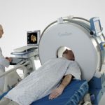
I recently had the opportunity to see a portable head CT scanner in action, the CeroTom by NeuroLogica (Danvers, MA). Today, I’ll give my thoughts on this new technology.

There are 3 major considerations when evaluating portable CT scanning:

  • Patient safety, always at the forefront
  • Usefulness, also know as image quality
  • Financial viability

From a safety standpoint, portable scanning can decrease (but not eliminate) the safety hazards associated with transporting a critically ill patient out of the ICU. Road trips are associated with misplaced/displaced lines, tubes and monitors about 15% of the time. These are lifelines in some patients, and even momentary disruptions can be life-threatening. Some patients are on levels of support so high they are not transportable, so portable scanners offer an opportunity to get diagnostic imaging that would not be available otherwise.

Clinical performance is on par with standard scanners. Resolution is lower, but the diagnostic accuracy and reliability are not different compared to fixed scanners.

From a financial standpoint, use of the portable scanner works as well. The Cleveland Clinic deployed a CereTom scanner a few years ago and found that the unit paid for itself in 6.9 months. For you financial types, the internal rate of return was 169% and the 5-year expected economic benefit was $2.6 million.

Bottom line: This new piece of technology offers significant benefits to patients in the ICU who may otherwise not be able to get imaging due to safety reasons. It can also be employed in the OR on anesthetized patients, which can assist with diagnosis in patients with both abdominal injuries requiring immediate operation and concomitant head injury.

Practical notes: The CereTom is an 8-slice scanner with a 25cm field of view. The patient is moved onto a scan board which supports the head while it is moved slightly off the top of the bed to accommodate the scanner. Current scanner cost is $450,000 and attachment packages for hospital beds are $7,000. One CT technologist can operate the unit, which takes about 5 minutes to set up and 15 minutes to scan. All lines, tubes and monitors must be (carefully) moved to the side of the bed so the scanner can fit over the top.  

References:

  • The economic and clinical benefits of portable head/neck CT imaging in the intensive care unit. Radiology Manage 30(2):50-54, 2008.
  • Review of portable CT with assessment of a dedicated head CT scanner. Am J Neuroradiol 30:1630-1636, 2009.

I have no financial interest in Neurologica, Inc.

Results – Blunt Trauma Radiographic Imaging Protocol

In my previous post (click here to view) I discussed an imaging protocol that we developed and implemented last year. Today, I’ll detail what it has accomplished in our patients.

We looked at 229 patients who had their imaging performed according to the new protocol during a 3 month period and compared them to 215 patients who were imaged the previous year. Each scan administered to each body area (head, chest, abdomen/pelvis, c-spine, t-spine, l-spine, face, neck angio) were tabulated separately.

We found that the overall number of scans performed decreased significantly. We looked at our data and generated numbers per 100 patients. During the control period, we did 298 CT scans per 100 patients. This decreased to 271 during the study period. The number of head scans remained the same (82 per 100 patients during control, 85 per 100 during the study), as did the cervical spine scans (84 vs 86).

The biggest declines were seen in chest CT (53 per 100 control vs 33 per 100 study) and abdominal CT (57 vs 43).

We did see an increase in conventional xrays of the thoracic and lumbar spines to offset the absence of reformatted spine images that would have been generated from the chest and abdominal CT scans. We also noted small increases in CT of the head, cervical spine, and neck angio. This was likely due to better adherence to specific guidelines.

Bottom line: we believe that our work shows that careful adoption of well thought out guidelines can make a difference in practice and significantly decreases radiation exposure in our blunt trauma patients.

To read the post on this protocol, or to download it, click here.

Click here to download the Blunt Trauma Radiographic Imaging Protocol Worksheet

Double CT Scanning in Trauma

Typical order: “chest CT with and without contrast”

A review of Medicare claims from 2008 showed that 5.4% of patients received double CT scans of the chest. Although the median was about 2% across 3,094 hospitals, 618 hospitals performed double scans on more than 10% of their patients. And 94 did it on more that half! One of the outliers was a small hospital in Michigan that double scanned 89% of Medicare patients! As expected, there was wide variation from hospital to hospital, and from region to region around the US. 

Time for some editorial comment.

This practice is very outdated and shows a lack of understanding of the information provided by CT. Furthermore, it demonstrates a lack of concern for radiation exposure by both the ordering physician and the radiologist, who should know better. 

Some officials at hospitals that had high scan rates related that radiologists ordered or okayed the extra scan because they believed that “more information was better.” There are two problems with this thinking.

  1. Information for information’s sake is worthless. It is only important if it changes decision making and ultimately makes a difference in outcome. 
  2. As with every test we do, there may be false positives. But we don’t know they are false, so we investigate with other tests, most of which have known complications. 

The solution is to do only what is clinically necessary and safe. The tests ordered should be based on the best evidence available, which demands familiarity with current literature.

In trauma, there are a few instances where repeat scanning of an area is required. Examples include solid organ lesions which may represent an injury or a hemangioma, and CT cystogram to exclude bladder trauma. In both cases, only a selected area needs to be re-scanned, not the entire torso.

Bottom line: Physicians and hospitals need to take the lead and rapidly adopt or develop guidelines which are literature-based. State or national benchmarking is essential so that we do not continue to jeopardize our patient’s safety and drive up health care costs.

Tomorrow I’ll share the blunt trauma imaging protocol we use which has decreased trauma CT use significantly at Regions Hospital.

Related posts:

Trauma Mortality vs Cancer Mortality from CT Scans for Trauma

Trauma professionals worry about radiation exposure in our patients. A lot. There are a growing number of papers dealing with this topic in the journals every month. The risk of dying from cancer due to CT scanning is negligible compared to the risk from acute injuries in severely injured patients. However, it gets a bit fuzzier when you are looking at risk vs benefit in patients with less severe injuries. Is it possible to quantify this risk to help guide our use of CT scanning in trauma?

A nice paper from the Mayo clinic looked at their scan practices in 642 adult patients (age > 14) over a one year period. They developed dose estimates using a detailed algorithm, and combined them with data from the Biological Effects of Ionizing Radiation VII data. The risk level for injury was estimated using their trauma team activation criteria. High risk patients met their highest level activation criteria, and intermediate risk patients met their intermediate level activation criteria.

Key points in this article were:

  • Average radiation dose was fairly consistent across all age groups (~25mSv)
  • High ISS patients had a significantly higher dose
  • Cumulative risk of cancer death from CT radiation averaged 0.1%
  • This risk decreased with age. It was highest in young patients (< 20 yrs) at 0.2%, and decreased to 0.05% in the elderly (> 60 yrs)

Bottom line: Appropriate CT scan use in trauma evaluation is challenging. It’s use is widespread, and although it changes management it has not decreased trauma mortality. This paper shows that the risk of death from trauma in the elderly outweighs the risk of death from CT scan radiation. However, this gap narrows in younger patients with less serious injuries because of their very low mortality rates. Therefore, we need to focus our efforts to reduce radiation exposure on our young patients with minor injuries.

Related posts:

References:

  • Comparison of trauma mortality and estimated cancer mortality from computed tomography during initial evaluation of intermediate-risk trauma patients. J Trauma 70(6):1362-1365, 2011.
  • Health risks from low levels of ionizing Radiation: BEIR VII, Phase 2. Washington DC: The National Academies Press, 2006.

Using CT To Diagnose Extremity Vascular Injury

The traditional gold standard for diagnosis of vascular injury to the extremities has been a good physical exam plus conventional catheter angiography. However, using angiography always adds a layer of complexity and risk to patient care. The interventional team may not be immediately available after hours, there is typically a road trip within the hospital to deliver the patient for the study, and overall it is quite expensive.

With the advancements we have seen in CT angio techniques and scanner technology, some centers have been using computed tomography to evaluate for vascular injury. A few small retrospective studies have been done, but this month a larger prospective study was published.

Over a 20 month period, 635 patients with extremity trauma and a suspicion for vascular injury were entered into the study. A structured physical exam was performed, and any patient with “hard signs” of vascular injury were taken to the OR. 527 patients had no signs of vascular injury and were observed and released. The remaining 73 (most had soft signs of vascular injury) underwent CT angiography of the extremity.

The sensitivity and specificity of this test were 82% and 92%, respectively. Positive and negative results were nearly perfectly predictive. However, approximately 10% were inconclusive, usually due to bullet artifact or reformatting errors. These patients either underwent confirmatory conventional angiography or operation.

Bottom line: Angiography using multi-detector CT scanners is an excellent tool for evaluating potential extremity vascular trauma from penetrating trauma. The technology is available around the clock without a wait, and usually does not involve lengthy trips through the hospital. A good physical exam is imperative so patients with hard signs of injury can go straight to the OR. Equivocal studies must be evaluated further by conventional angio or an operation.

Reference: Prospective multidetector computed tomography for extremity vascular trauma. J Trauma 70:808-815, 2011.