Tag Archives: ct scan

Extraperitoneal Bladder Rupture

This injury is likely to occur in patients who have a full bladder and sustain anterior pelvic trauma that typically leads to fractures. They generally present with gross hematuria upon placement of the bladder catheter. This should prompt an abdominal CT scan with cystogram technique.

CT cystogram involves pressurizing the bladder with contrast prior to the study. This differs from the usual method of clamping the catheter and allowing the bladder to passively fill. The literature here is clear: failure to use cysto technique will miss 50% of these injuries.

The majority of extraperitoneal bladder injuries can be treated nonoperatively, and probably do not need Urology involvement. The bladder catheter is left in place 10-14 days (we do 10 days), and a repeat cystogram is obtained. If there is no leak, the catheter can be removed. If there is still some leakage, Urology consultation should then be obtained. 

There are a few cases where operative management is required:

  • There is some intraperitoneal component of bladder injury
  • Fixation of the pubic rami is required (bathing the orthopedic hardware with urine is frowned upon)
  • Failure of conservative management

Arrows in the photo show extraperitoneal extravasation of cystogram contrast.

Related posts:

Trauma 20 Years Ago: CT Imaging of the Aorta

CT scan is now the standard screening test for injury to the thoracic aorta. But 20 years ago, we were still gnashing our teeth about how to detect this injury.

An interesting paper was published in the Journal of Trauma 20 years ago this month on this topic. Over a 2 year period, the Medical College of Wisconsin at Milwaukee looked at all patients who underwent imaging for aortic injury. At the time the gold standard was aortogram. They looked at patients who underwent this study and CT, which was not very common at the time.

They had 50 patients who underwent aortography alone and 17 who underwent both tests. Of the 17, 5 had the injury, but only three were seen on CT. There were also two false positives. Sensitivity was 83%, specificity was 23%, with 53% accuracy. The authors concluded that any patients with strong clinical suspicion of aortic injury should proceed directly to aortogram.

Why the difference today? Scan technology and resolution has increased immensely. Also, the timing of IV contrast administration has been refined so that even subtle intimal injuries can be detected. CT scan is now so good that we have progressed from the CV surgeon requiring an aortogram before they would even consider going to the OR, to the vascular surgeon / interventional radiologist proceeding directly to the interventional suite for endograft insertion.

Radiation Exposure in Pediatric Trauma

The use of radiographic imaging in trauma patients has exploded over the past decade. A growing amount of research is looking at adult patients, but what about children?

Johns Hopkins did a one year retrospective review of radiographic imaging in kids age 14 and below. The studies performed and the estimated radiation dose was calculated for each child. A total of 719 children were studied and they underwent a total of 4603 studies:

  • CT scans – 1457 (32%)
  • Plain radiographs – 3097 (67%)
  • Fluoroscopy – 49 (1%)

CT accounted for only 32% of studies but delivered 91% of the total radiation dose. Children involved in car crashes received the highest dose of radiation (18mSv) versus burned children, who had the lowest dose (1.2 mSv). Radiation exposure increased as the injury severity increased. The average age was 8 years, which means that these children have a long time until possible side-effects emerge.

What to do? First, seriously weigh the risks and benefits of every radiographic study before you order it. If CT is not essential, do something else. The ALARA concept is key (as low as reasonably achievable):

  • Use weight-based CT protocols in order to deliver the minimum amount of radiation needed to get decent images
  • Shield all sensitive areas that are not being imaged
  • Use focused studies
  • Avoid repeat exams
  • Become knowledgeable about the effects of radiation exposure
  • Ask yourself: “What if this were my child?

Reference: Brown, et al. Diagnostic radiation exposure in pediatric trauma patients. J Trauma 2010, ahead of print. 

How Often Are Imaging Studies Repeated After Trauma Transfers?

Smaller trauma hospitals, both designated and undesignated, are the front line for the initial care of the majority of trauma patients. Many patients can be evaluated and sent home or admitted to the initial hospital. More severely injured patients are commonly transferred to the nearest Level I or Level II trauma center for care of injuries requiring specialists.

Imaging studies such as conventional xray and CT scan are a necessary part of the initial trauma evaluation. But is it necessary to do a full radiographic evaluation, even when it is known that the patient will have to be transferred?

Researchers at Dartmouth Hitchcock Medical Center examined the issue of repeat imaging at their Level I center. They looked at 138 patients that were transferred to them from other rural hospitals. They found that 75% underwent CT scanning prior to transfer, and 58% underwent repeat scanning upon arriving at Dartmouth.

The authors discovered the following:

  • Head CTs were repeated 52% of the time, primarily due to clinical indications
  • Spine reconstructions were repeated 33-50% of the time due to inadequate reconstruction technique
  • Chest (31%) and abdomen (20%) were repeated due to inappropriate use of IV contrast
  • 13% of image disks used incompatible software
  • 7% of images were not sent with the patient

Here are my recommendations for imaging by hospitals that refer patients to Level I or II trauma center:

  • Obtain the essential plain films recommended by ATLS (chest, pelvis)
  • If an obvious injury requiring transfer is found on exam (e.g. open fracture) do no further studies
  • Obtain any imaging studies needed to decide if you can admit the patient to your own hospital (example: abdominal CT for abdominal pain and negative FAST. Keep if no injury, transfer if solid organ injury)
  • As soon as an injury is identified that mandates transfer, do no further studies
  • Always send image disks with the patient
  • Work with your referral trauma center to obtain a copy of their CT imaging protocols so if you do need to perform a study you can duplicate their technique

Reference: Gupta et al. Inefficiencies in a Rural Trauma System: The Burden of Repeat Imaging in Interfacility Transfers. J Trauma 69(2):253-255, 2010.

Cervical Spine Clearance in Obtunded Patients

Cervical spine clearance in obtunded trauma patients has always been controversial. Most physicians believe that evaluation of bones and ligaments is required, although there is a minority that say that the spine can be cleared purely by radiographs. This would greatly simplify the process and decrease costs.

A prospective study was presented at EAST in January that evaluated the use of CT alone to clear the c-spine in these patients. It was presented by Claridge et al from MetroHealth in Cleveland, and is an expansion of an earlier prospective they performed. Based on the original study, the protocol was revised and the results of this re-study was presented.

The study involved 197 patients who were victims of blunt trauma, obtunded, and were noted to move all extremities. Short term mortality was 13% and long term mortality was 27%, which shows how badly injured this group was. The average ISS was 23 and the initial GCS was 8.

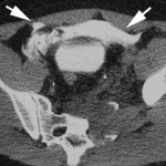
The following radiographic criteria were used to diagnose a significant c-spine injury:

  • Fracture line extending on 2 consecutive CT slices
  • Marked prevertebral soft tissue swelling or hematoma
  • Malalignment not explained by degenerative changes
  • Abnormal facets or posterior malalignment on sagittal reconstruction
  • Occipital condyle injury involving the craniocervical junction

Followup was performed either by re-examination after awakening (62%), followup by phone or chart review (12%), or MRI for persistent c-spine pain (2%). Thirteen percent died before re-evaluation, and 11% were lost to followup.

Using this protocol, the average hospital day of clearance decreased from 7.5 to 3.3, the incidence of decubitus ulcer from the collar decreased from 5% to 0.5%, and the average length of stay decreased from 23 to 14 days. All of these results were statistically significant.

The authors recognized that long term followup was lacking in this study and there was the potential for missed injury. Power calculations show that there are not enough patients enrolled to give a statistically sound result. The issue of spinal cord injury without radiographic abnormality (SCIWORA) is always a possibility.

The bottom line: clearance based on radiographs alone is still not ready for prime time. Some injuries will ultimately be missed, and a fraction of those can cause devastating injury. The real question to be answered is “How many missed injuries is okay?” Until more and better work is done, some combination of radiographic and clinical techniques must be used.

Reference: A normal CT alone may clear the cervical spine in obtunded blunt trauma patients with gross extremity movement – a prospective evaluation of a revised protocol. Claridge et al, MetroHealth Medical Center. Presented at the 23rd Annual Scientific Assembly of the Eastern Association for the Surgery of Trauma, January 2010.