Tag Archives: complications

Consequences Of Embolizing Renal Injuries

In my last post, I noted that nonoperative management is the norm for dealing with high grade renal injuries. One of the possible options, angioembolization, was relatively infrequently used at only 6% of the time.

For management of other organs like the spleen, there are several angioembolization options. Depending on the type and severity of injury, selective (partial) or nonselective (main splenic artery) embolization can be carried out. For the liver, only selective embolization can be used. But what about the kidney? 

Are there consequences of nonselective renal embolization? Or should we always strive for selective control? The urology group of the University of Tennessee – Knoxville published a series of papers on their experience using embolization in patients with the most severe injuries (Grade 5). They retrospectively examined just over 3 years of admissions with this injury. Numbers were very small (6 men, 3 women).

But they also published a second paper, extending the review dates to capture one more male patient. And they followed this group for 1.5 to 5 years (mean 2.5 years) to determine if any delayed complications surfaced.

Here are the factoids:

  • Seven patients underwent full, nonselective embolization, and the other three had “super selective” embolization
  • All patients had control of bleeding without surgical intervention
  • Followup CT imaging showed no persistent extravasation or expanding hematoma
  • No patient developed complications, such as a retroperitoneal abscess, prolonged fever, or hypertension while in the hospital or during short-term followup
  • Most patients showed a very small increase in serum creatinine (mean 0.04), but one patient increased from 1.1 to 1.7
  • On longer term followup, one patient, age 51, developed hypertension 10 months after his injury. It is not possible to determine whether he was one of the 20% of older adults who develop hypertension, or whether it was due to the procedure. it was well-controlled with a single antihypertensive med.
  • None developed altered renal function, stones, chronic pain, fistula, or pseudoaneurysm

Bottom line: Obviously, the data is very limited with only 10 patients. However, it is very interesting to note that the majority of these patients underwent nonselective embolization of the renal artery without any adverse event. The one case of hypertension occurred with nonselective embolization, although I have seen several case reports where this occurs with selective embolization as well.

It is now well-accepted that high-grade renal injury can and should be managed nonoperatively if the patient’s hemodynamic status is reasonable. I recommend a trip to interventional radiology if the patient has active extravasation or a high-grade (Grade 4 or 5) injury, as these patients are at risk for loss of the entire kidney otherwise. Selective embolization can be attempted first, but don’t be shy to take out the entire organ if need be. 

References: 

  • Percutaneous embolization for the management of Grade 5 renal trauma in hemodynamically unstable patients: initial experience. J Urology 181:1737-1741, 2008.
  • Intermediate-term follow-up of patients treated with percutaneous embolization for Grade 5 blunt renal trauma. J Trauma 69(2):468-470, 2010.

Top 10 Worst Complications: #1 Nasocerebral Tube

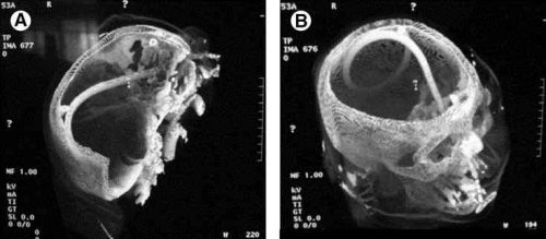
Minor complications from nasogastric tube insertion occur relatively frequently. Emesis is fairly common when the gag reflex is stimulated by the tube in the back of the oropharynx. An infrequent but possibly fatal one is insertion through the cribriform plate. 

The cribriform plate is located directly posterior to the nares and is part of the ethmoid bone. It is very porous in nature and weaker than the surrounding portions of the ethmoid. It is easily fractured, and can be seen is association with basilar skull fractures. This is one source for rhinorrhea in patients with these fractures.

Cribriform fracture is a contraindication to unprotected insertion of a nasogastric tube. If you look at the sagittal section below, the plate lies directly behind the nares. When inserting the NG tube, we are usually taught to aim the tube straight back. Unfortunately, this aims it directly at the cribriform. If a fracture is present, it is possible that you may be inserting a nasocerebral tube!

Cribriform plate - sagittal section

The usual symptoms when this occurs consist of immediate neurologic deterioration to coma, and a unilateral or bilateral blown pupil. The tube must not be withdrawn, because it will cause significant injury to the base of the brain. A stat neurosurgical consultation must be obtained, and if the patient is salvageable, the tube must be withdrawn through a craniectomy.

To avoid this dreaded complication, identify patients at risk for cribriform injury. They are:

  • patients with signs of trauma from eyebrows to zygoma
  • comatose patients
  • patients with signs of basilar skull fracture (Battle’s sign, raccoon eyes, oto- or rhinorrhea)

If your patient is at risk, follow these guidelines:

  • first, does the patient really need a gastric tube?
  • if comatose, insert an orogastric tube
  • if awake, don’t put the tube in their mouth, as they will gag continuously. Instead, place a lubricated, curved nasal airway. Then lube up a slightly smaller Salem sump tube and pass it through the airway.

Air Embolism From an Intraosseous (IO) Line

Intraosseous (IO) lines are a godsend when we are faced with a patient who desperately needs access but has no veins. The tibia is generally easy to locate and the landmarks for insertion are straightforward. They are so easy to insert and use, we sometimes “set it and forget it”, in the words of infomercial guru Ron Popeil.

But complications are possible. The most common is an insertion “miss”, where the fluid then infuses into the knee joint or soft tissues of the leg. Problems can also arise when the tibia is fractured, leading to leakage into the soft tissues. Infection is extremely rare.

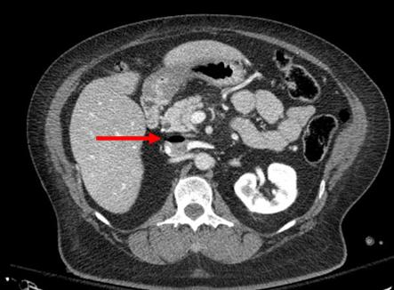
This photo shows the inferior vena cava of a patient with bilateral IO line insertions (black bubble at the top of the round IVC).

During transport, one line was inadvertently disconnected and probably entrained some air. There was no adverse clinical effect, but if the problem is not recognized and the line is not closed properly, there could be.

Bottom line: Treat an IO line as carefully as you would a regular IV. You can give anything through it that can be given via a regular IV: crystalloid, blood, drugs. And even air, so be careful!

Incidental Finding: Gas In The Spleen After Embolization

Most solid organ injury practice guidelines include angioembolization in part of the pathway. But very few require re-imaging at any point to see how the liver or spleen are coming along.

But every once in a while another condition arises, or symptoms worsen unexpectedly, causing us to get another CT scan that includes the abdomen and pelvis. And sometimes we see things that we wouldn’t normally see, like air bubbles in the organ that was embolized.

So what is okay, and what requires some kind of intervention? Our friends at ShockTrauma in Baltimore looked at this in 2001 and can provide some pretty good guidance. They reviewed patients who underwent CT scan both before and after embolization over about 2.5 years. They performed the post-embolization scans for specific indications like fevers, elevated WBC count (!), increasing abdominal pain, or an episode of hypotension. A total of 53 patients were studied.

Here are the factoids:

  • 24 patients underwent embolization of the main splenic artery, 22 had selective embolization of part of the spleen, and 7 had both
  • Splenic infarcts occurred in 63% of patients with main artery embolization, but were large (> 50% of the parenchyma) in only 20% of those
  • Infarcts occurred in 100% of selective embolizations, but were small (< 50%) in 93% of cases
  • Infarcts occurred in 71% of patients with both main and selective embolization, and most were small (80%)
  • Seven (13%) patients developed gas bubbles in the spleen, and was usually present for 1-7 days before disappearing
  • One patient developed increasing gas with pneumoperitoneum and underwent splenectomy for a splenectomy for abscess

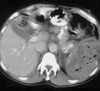
This picture that shows tiny bubbles in the spleen parenchyma that represent “normal” gas after embolization:

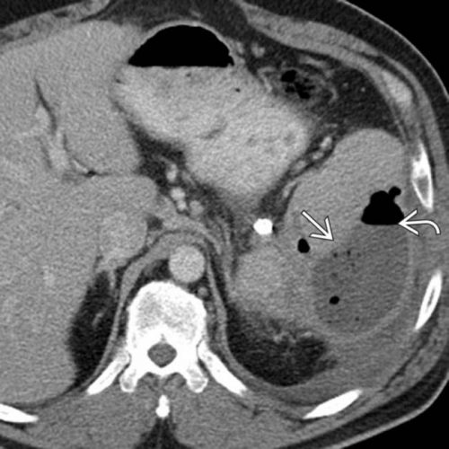
And the following one shows an air/fluid collection in the spleen that indicates an abscess:

Bottom line: Tiny bubbles in the spleen (and probably the liver) occur normally after angioembolization. They usually develop within an area of infarction, and most are benign. It is possible for them to evolve into a splenic abscess, but unlikely. Many embolization patients develop fevers at some point, and most have an elevated WBC count. So in most cases, you can ignore this incidental finding, as long as your patient has mild symptoms.

However, if the patient develops high fevers, very elevated WBC (> 25K), increasing abdominal or flank pain, and the spleen develops an air/fluid level, an abscess is forming. Despite what your radiologist might suggest, catheter drainage is not a good idea. The tubes are too small to remove the slurry that is generally found within the abscess. A trip to the OR is the only effective treatment, and splenectomy is generally the only option.

Related posts:

Reference: CT Findings after Embolization for Blunt Splenic Trauma. J Vasc Intervent Radiol 12(2):209-214, 2001.

EAST 2017 #8: When Is “Mild TBI” Not So Mild?

Traumatic brain injury (TBI) is very common, with the majority falling into the “mild” category. This is usually defined as patients with injury to the head and a GCS of 13-15. These uncomplicated patients are frequently discharged from the emergency department, or undergo only a brief evaluation if admitted for other reasons.

The group at Shock Trauma focused on a less appreciated subset of mild TBI patients, those whose condition is a little more complicated. Specifically, these are patients with GCS 13-14 with positive findings on head CT leading to a calculated abbreviated injury score (head) of > 2, and some persistence of their symptoms while in the hospital. At many hospitals (including my own), these patients receive an inpatient TBI evaluation. But if they pass this initial screening, they are not consistently referred for any outpatient TBI followup.

Are these mild, complicated TBI patients (mcTBI) unique? Do they behave the same as the uncomplicated ones? The research group performed a prospective study on patients who sustained an mcTBI over a 4 month period.  They excluded patients with mental illness, dementia, and non-English speaking and homeless patients. They tried to contact patients up to three times after discharge to administer several standard tests and determine if they had any specific residual symptoms.

Here are the factoids:

  • Of the 142 patients with mcTBI during the study period, there was substantial attrition over time, with only 25 remaining at 6 months and 10 at one year
  • 64% of patients who responded at 6 months remained symptomatic. Depression, dizziness, and a feeling of impaired health were common.
  • 80% of patients still described symptoms at one year. The same complaints were most common, and some required changes in activities of daily living or assistive devices.

Bottom line: Although small and fraught with the usual problems in long-term tracking of urban trauma patients, this study is eye-opening. We too often dismiss “mild TBI” and being almost nothing, even in patients with positive findings on head CT. This work suggests that we are underestimating the needs of those patients. The authors used this data to design longer-term care processes for this subset of patients. Other centers should follow suit to make sure these patients’ post-injury needs are better met.

Questions and comments for the authors/presenters:

  • Describe the possible biases that patient selection and attrition may have had on the study
  • What type of TBI screening do you use in the hospital?
  • Given that a number of assessments were administered over the phone, I look forward to hearing some of the other details not listed in the abstract
  • Was there any correlation between specific CT findings and later symptoms?
  • Provide details of your long-term care programs for these patients
  • I enjoyed this thought provoking abstract!

Click here to go the the EAST 2017 page to see comments on other abstracts.

Related posts:

Reference: Mild TBI is not ‘mild’… survivors tell their complicated stories. Quick Shot #3, EAST 2017.