Tag Archives: complications

Complications of Splenic Embolization for Trauma

Angioembolization has become a common procedure that can increase the likelihood of success for nonoperative management for splenic trauma. It does have its own set of complications to be aware of, however.

The most obvious complication is mechanical injury to the femoral artery. This occurs in 1 to 3% of patients. It is more common in the very young (small caliber artery) and the elderly (arteries of stone). Rarely, the substance or device that is used for the embolization may migrate or end up on the wrong spot, infarcting something important.

A common issue that occurs is infarction of portions of the spleen. This is actually the desired effect, as it stops the bleeding. Most of the time, we are unaware of the changes that take place in the spleen post-procedure. But every once in a while we get a repeat CT scan days or weeks down the road and see some very interesting things.

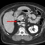
The most common finding is a splenic infarct alone. This is an area of the spleen, sometimes wedge shaped, that does not take up contrast. This is normal. In some cases, gas bubbles are seen within the spleen parenchyma, usually within the infarcted area. In others, large areas of gas are present, and an air-fluid level may also be seen. This is definitely not normal.

Note the infarcted area at the arrow, with a tiny gas bubble visible.

Tiny bubbles are normal after this procedure, and can be ignored if the patient does not appear ill and does not have any systemic evidence of inflammation or sepsis. On the other hand, big bubbles or air-fluid levels probably indicate a developing splenic abscess, and the patient will usually appear ill and have a high WBC count. Unfortunately, the only treatment for this is splenectomy. Insertion of drainage catheters does not work and the patient will only become sicker if it is attempted.

Best Of: VIP Syndrome In Healthcare (Very Important Person)

The VIP syndrome occurs in healthcare when a celebrity or other well-connected “important” person receives a level of care that the average person does not. This situation was first documented in a paper published in the 1960s which noted that VIP patients have worse outcomes.

VIPs have the expectation that they can get special access to care and that the care will be of higher quality than that provided to others. Healthcare providers often grant this extra access, in the form of returned phone calls and preferential access to their clinic or office. The provider tries to provide a higher quality of care by ordering additional tests and involving more consultants. This idea ignores the fact that we already provide the best care we know how, and money or fame can’t buy any better.

Unfortunately, trying to provide better care sets up the VIP for a higher complication rate and a greater chance of death. Healthcare consists of a number of intertwined systems that, in general, have found their most efficient processes and lowest complication rates. Any disturbance in this equilibrium of tests, consultants, or nursing care moves this equilibrium away from its safety point.

Every test has its own set of possible complications. Each consultant feels compelled to add something to the evaluation, which usually means even more tests, and more possible complications. And once too many consultants are involved, there is no “captain of the ship” and care can become fragmented and even more inefficient and dangerous.

How do we avoid the VIP Syndrome? First, explain these facts to the VIP, making sure to impress upon them that requesting or receiving care that is “different” may be dangerous to their health. Explain the same things to allproviders who will be involved in their care. Finally, do not stray from the way you “normally” do things. Order the same tests you usually would, use the same consultants, and take control of all of their recommendations, trying to do things in your usual way. This will provide the VIP with the best care possible, which is actually the same as what everybody else gets.

Reference: “The VIP Syndrome”: A Clinical Study in Hospital Psychiatry. Weintraub, Journal of Mental and Nervious Disease, 138(2): 181-193, 1964.

Delayed Diagnosis of Blunt Intestinal Injury in Children

Yesterday, I wrote about using ultrasound in place of CT for initial diagnosis of blunt abdominal injury in children. Although it looks good for identification of solid organ injury and free fluid, it may miss injury to the intestine. Is that bad?

Lets look at a recent study that examined the consequences of delayed laparotomy for blunt intestinal injury. The American Pediatric Surgical Association conducted an 18-center study of the management of intestinal injuries in children less than 16 years of age. They were stratified by time to treatment. There were 214 patients with complete data records for review. 

The majority of the patients were involved in a motor vehicle crash or a bicycle accident. Demographics were similar in all time to treatment groups. Half were resuscitated at a referring hospital and then transferred to a pediatric trauma center, on average after 6 hours.

Key points:

  • The only deaths occurred in the 0-6hr and 6-12hr groups. The average Injury Severity Score of the children who died was significantly higher than survivors.
  • Children operated on in the 0-6hr group had significantly higher ISS as well.
  • There was no difference in early or late complications across all groups.
  • Time to beginning oral intake and time in hospital were the same in all groups.

The authors concluded that observation and serial exam rather than urgent exploration or repeated CT scans is appropriate.

Bottom line: If you combine this study with the ultrasound study I reviewed yesterday, it seems appropriate to modify the usual (read: adult) way of evaluating blunt trauma to the abdomen. In place of automatically getting a CT scan of the abdomen in children, obtain a complete abdominal ultrasound to detect solid organ injury or free fluid. This will determine the degree of monitoring needed (e.g. ICU for higher grade liver or spleen injuries). Follow this with serial abdominal exam. If the child becomes symptomatic, it’s probably time to proceed to the OR. Note: I generally do not make children npo during the observation phase. They need to eat, and if they don’t want to, that tells you something.

Related post: Sonography in pediatric abdominal trauma

Reference: Delay in diagnosis and treatment of blunt intestinal injury does not adversely affect prognosis in the pediatric patient. J Pediatric Surg 45(1):161-166, 2010.

Less Morbidity From Negative Trauma Laparotomies?

Trauma surgeons generally dread the negative laparotomy for trauma. Previous work has shown that complications occur in anywhere from 22% to 53% of cases. Those studies were usually retrospective and included patients with penetrating trauma, which may have skewed the results.

A newly published study tries to throw this common wisdom in doubt. It was a retrospective review of a prospectively maintained database of trauma admissions after blunt trauma . Patients were separated into groups who underwent immediate, delayed or no laparotomy, as well as whether they had or did not have associated injuries. Complications were tracked using an accurate and validated tracking system. The complications tracked included death, DVT, PE, infections, pulmonary issues, as well as other organ system problems.

The authors found that a negative laparotomy did not increase the complication rate, but that a delayed laparotomy did. They also noted that a Complication Impact Score (that they made up) was higher in the delay to laparotomy patients. So they believe that when clinical and imaging findings are equivocal, doing an operation to establish a diagnosis is justifiable.

My Bottom Line: This study does not look at really delayed complications like small bowel obstruction, which we see with some regularity in old trauma patients. Also, other studies have also shown that brief observation, even in patients with a bowel injury, does not increase complications significantly. Unless the potential injury that you are observing is known to have significant complications, my practice is to observe equivocal cases in order to avoid more complications down the road.

Reference: “Never be wrong”: the morbidity of negative and delayed laparotomies after blunt trauma. J Trauma 69(6): 1386-1392, 2010.

Air Embolism From an Intraosseous (IO) Line

IO lines are a godsend when we are faced with a patient who desperately needs access but has no veins. The tibia is generally easy to locate and the landmarks for insertion are straightforward. They are so easy to insert and use, we sometimes “set it and forget it”, in the words of infomercial guru Ron Popeil.

But complications are possible. The most common is an insertion “miss”, where the fluid then infuses into the knee joint or soft tissues of the leg. Problems can also arise when the tibia is fractured, leading to leakage into the soft tissues. Infection is extremely rare.

This photo shows the inferior vena cava of a patient with bilateral IO line insertions (black bubble at the top of the round IVC). During transport, one line was inadvertently disconnected and probably entrained some air. There was no adverse clinical effect, but if the problem is not recognized and the line closed, there could be.