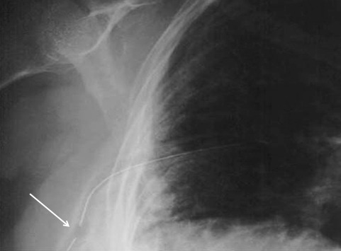
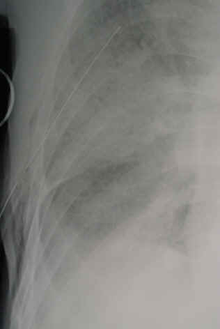
Tag Archives: chest tube

What To Do? Chest Tube Repositioning

It happens from time to time. Your patient has a hemothorax or pneumothorax and you insert a chest tube. Well done! But then the xray comes back:

The last hole in the drain is outside the chest! What to do???

Here are the questions that need to be answered:

  • Pull it out, leave it, or push it in?
  • Does length of time the tube has been in make a difference?
  • Does BMI matter?

Leave comments below regarding what you do. Hints tomorrow and answers on Monday!

Chest Tube Management Protocol – Pediatric

Yesterday I described a protocol for deciding when to remove a chest tube in adults. Today, I’ll go over a variant of this algorithm for children. In general, it’s very similar. The major change is in the volume criterion. In adults, we decided upon an (arbitrary) value of 150cc per three shifts. We chose a time interval of 3 shifts vs 1 day to speed up the process.

Suppose you use the 1 day rule for looking at chest tube output. Typically, this would be evaluated in the morning, and the process of pulling the tube or applying water seal, followed by delayed xrays, could lead to a very late discharge. If the output is checked every shift and the most recent three are summed, the patient could meet criteria later in the day and have the tube pulled in the evening. This would allow for an earlier discharge the following day, shaving 12 hours or more off of the hospital stay. This may not make much of a difference to the hospital (although for busy ones it does), but it’s huge for patient comfort and satisfaction.

Click this image or the link below for a full-size version.

Note that the output criterion has been changed to 2cc/kg over three shifts. This adjusts for the varying sizes of the children that we treat. Otherwise things are basically the same.

Related posts:

Chest Tube Management Protocol – Adult

Chest tubes are needed occasionally to help manage chest injuries. How do you decide when they are ready for removal?

Unfortunately, the literature is not very helpful in answering this question. To come up with a uniform way of pulling them, our group looked at any existing literature and then filled in the blanks, negotiating criteria that we could all live with. We came up with the following. Click the image to see a full-size version, or click the link below.

Removal criteria:

  • No (or a minimal, stable) residual pneumothorax
  • No air leak
  • Less than 150cc drainage over the past 3 shifts. We do not use daily numbers, as it may delay the removal sequence. We have moved away from the “only pull tubes on the day shift” mentality. Once the criteria are met, we begin the removal sequence, even in the evening or at night.

Removal sequence:

  • Has the patient ever had an air leak? If so, they are placed on water seal for 6 hours and a followup AP or PA view chest x-ray is obtained. If no pneumothorax is seen, proceed to the next step. If there was no air leak, skip this step.
  • Pull the tube. Click here to see a video demonstrating the proper technique.
  • Obtain a followup AP or PA view chest x-ray in 6 hours.
  • If no recurrent pneumothorax, send the patient home! (if appropriate)

Click here to download the full printed protocol.

Pigtail Catheters Instead Of Chest Tubes?

Traditionally, hemothorax and pneumothorax in trauma has been treated with chest tubes. I’ve previously written about some of the debate regarding using smaller tubes or catheters. A paper that will be presented at the EAST meeting in January looked at pain and failure rates using 14Fr pigtail catheters vs 28Fr chest tubes.

This was a relatively small, prospective study, and only 40 of 74 eligible patients were actually enrolled over 20 months at a Level I trauma center in the US. Pain was measured using a standard Visual Analog Scale, as was complication and failure rate, tube duration and hospital stay.

The following interesting findings were noted:

  • Chest wall pain was similar. This is expected because the underlying cause of the pneumothorax, most likely rib fractures, is unchanged.
  • Tube site pain was significantly less with the pigtail
  • The failure rate was the same (5-10%)
  • Complication rate was also the same (10%)
  • Time that the tube was in, and hospital stay was the same

Bottom line: There may be some benefit in terms of tube site pain when using a smaller catheter instead of a chest tube. But remember, this is a very small study, so be prepared for different results if you try it for your own trauma program. If you do choose to use a smaller tube or catheter, remember to do so only in patients with a pure pneumothorax. Clotted blood from a hemothorax will not be completely evacuated.

Related posts:

Reference: A prospective randomized study of 14-French pigtail catheters vs 28F chest tubes in patients with traumatic pneumothorax: impact on tube-site pain and failure rate. EAST Annual Surgical Assembly, Oral paper 12, 2013.

Retained Hemothorax And Empyema

Patients with chest trauma sustain hemothorax on occasion. The trauma professional usually picks this diagnosis up in the initial evaluation and makes a decision whether or not to drain it. The parameters for this decision are not very clear, even today. But what happens when there is residual hemothorax? Should we be more aggressive in getting it out?

All this boils down to an understanding of the natural history of retained hemothorax. This kind of information can help us decide whether to be more aggressive in our efforts to remove it. The results of a multicenter study looking at this issue was published recently. They focused on patients who had a chest tube placed for management of either hemo- or pneumothorax within 24 hours of admission. Patients who had suspected retained hemothorax after tube removal received a CT scan within 14 days. The usual outcomes were studied (length of stay, complications) as well as development of empyema (purulence, acidic pleural fluid, positive Gram stain or culture).

Some interesting results:

  • 328 patients were enrolled across 20 centers. Not a lot, but one of the bigger studies to date.
  • Empyema was diagnosed in 27% of patients
  • Risk factors identified included rib fractures, ISS > 25, and performance of additional interventions for drainage
  • Patients who developed empyema stayed in the ICU and the hospital longer

Bottom line: Retained hemothorax turns into a very serious problem in a quarter of trauma patients who have a chest tube inserted. The presence of residual blood after the chest tube is removed should prompt us to figure out if it’s solid clot or liquid blood (remember the old decubitus view chest xray? They still work!). If it’s liquid, consider drainage via thoracentesis or a smaller catheter. If it’s clot, it may require more invasive techniques to drain it (VATS). If you decide to send the patient home, have them watch out for fevers, chest pain, dyspnea and other symptoms and signs of a developing complication, and make sure they report it to you promptly.

Related post:

Reference: Development of posttraumatic empyema in patients with retained hemothorax: Results of a prospective, observational AAST study. J Trauma 73(3):752-757, 2012.