Tag Archives: chest tube

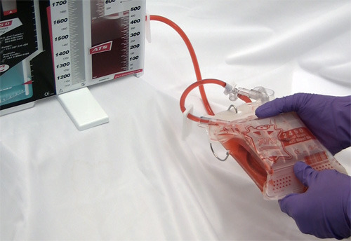
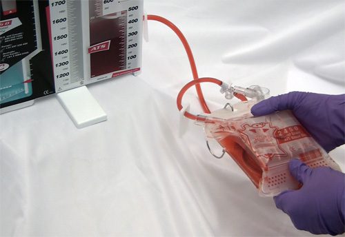
Autotransfusing Blood Lost Through The Chest Tube

Autotransfusing blood that has been shed from the chest tube is an easy way to resuscitate trauma patients with significant hemorrhage from the chest. Plus, it’s usually not contaminated from bowel injury and it doesn’t need any fancy equipment to prepare it for infusion. 

It looks like fresh whole blood in the collection system. But is it? A prospective study of 22 patients was carried out to answer this question. A blood sample from the collection system of trauma patients with more than 50 cc of blood loss in 4 hours was analyzed for hematology, electrolyte and coagulation profiles.

The authors found that:

  • The hemoglobin and hematocrit from the chest tube were lower than venous blood (Hgb by about 2 grams, Hct by 7.5%)
  • Platelet count was very low in chest tube blood
  • Potassium was higher (4.9 mmol/L), but not dangerously so
  • INR, PTT, TT, Factor V and fibrinogen were unmeasurable

image

Bottom line: Although shed blood from the chest looks like whole blood, it’s missing key coagulation factors and will not clot. Reinfusing it will boost oxygen carrying capacity, but it won’t help with clotting. You may use it as part of your massive transfusion protocol, but don’t forget to give plasma and platelets according to protocol. This also explains why you don’t need to add an anticoagulant to the autotransfusion unit prior to collecting or giving the shed blood!

Related post: Chest tubes and autotransfusion

Reference: Autotransfusion of hemothorax blood in trauma patients: is it the same as fresh whole blood? Am J Surg 202(6):817-822, 2011.

Pneumothorax: How Big Is Too Big?

One of the big unanswered questions in the management of pneumothorax is, how big is too big? At what size is a chest tube of some type mandatory? 

The problem is that we just don’t have any good data. Seems like a simple problem, right? Unfortunately, it’s not. A pneumothorax is a three dimensional collection that surrounds the lung in very random ways. All we had to detect and “measure” them for decades was the lowly chest x-ray. Unfortunately, this is a 2D shadow picture that does not allow us to accurately estimate the size.

A few old papers exist that tried to quantify pneumothorax, but they are of no use now that we have chest CT. Unfortunately this new technology has drawbacks, as well. First, it’s just a stack of 2D images that our minds assemble into a 3D mental model, so it’s still difficult to quantify the air. And second, you shouldn’t be getting a chest CT just to diagnose pneumothorax. In blunt trauma, it’s really just for checking the thoracic aorta for injury.

So we’re left with the original question, and there are three answers. If there is any physiologic compromise (hypoxia, tachypnea, anxiety), then the chest should be drained. If the pneumothorax is enlarging over serial chest x-rays, then it should be drained before it causes physiologic change. And finally, if there is concern that it is so large that it will take too long to absorb, especially in older patients with comorbidities, a chest drain should be inserted. This is a somewhat soft indication, however.

Bottom line: The three reasons above are the usual answers to the question, “how big is too big?” For me, once the pneumothorax pushes the lung 1-2 cm away from the chest wall from apex to base, it’s time for a tube.

Related posts:

Pigtail Cathers Instead Of Chest Tubes?

I reviewed this abstract a few months ago, and now I’ve had the opportunity to hear it and see the data. Here’s an update on whether this is worthwhile..

This was a relatively small, prospective study, and only 40 of 74 eligible patients were actually enrolled over 20 months at a Level I trauma center in the US. Pain was measured using a standard Visual Analog Scale, as was complication and failure rate, tube duration and hospital stay.

The following interesting findings were noted:

  • Chest wall pain was similar. This is expected because the underlying cause of the pneumothorax, most likely rib fractures, is unchanged.
  • Tube site pain was significantly less with the pigtail
  • The failure rate was the same (5-10%)
  • Complication rate was also the same (10%)
  • Time that the tube was in, and hospital stay was the same

There were a few questions regarding blinding of the pain scale raters, but other than the small sample size, the study was nicely done.

Bottom line: There may be some benefit in terms of tube site pain when using a smaller catheter instead of a chest tube. But remember, this is a very small study, so be prepared for different results if you try it for your own trauma program. If you do choose to use a smaller tube or catheter, remember to do so only in patients with a pure pneumothorax. Clotted blood from a hemothorax will not be completely evacuated.

Related posts:

Reference: A prospective randomized study of 14-French pigtail catheters vs 28F chest tubes in patients with traumatic pneumothorax: impact on tube-site pain and failure rate. EAST Annual Surgical Assembly, Oral paper 12, Jan 17, 2013.

What To Do? Chest Tube Repositioning

So you’re faced with a chest tube that “someone else” inserted, and the followup chest xray shows that the last drain hole is outside the chest. What to do?

Well, as I mentioned, there is very little written on this topic, just dogma. So here are some practical tips on avoiding or fixing this problem:

  • Don’t let it happen to you! When inserting the tube, make sure that it’s done right! I don’t recommend making large skin incisions just to inspect your work. Most tubes can be inserted through a 2cm incision, but you can’t see into the depths of the wound. There are two tricks: 
  1. In adults with a reasonable BMI, the last hole is in when the tube markings show 12cm (bigger people need bigger numbers, though)
     
  2. After insertion, get into the habit of running a finger down the radiopaque stripe on the tube all the way to the chest wall. If you don’t feel a hole (which is punched through the stripe), this will confirm that the it is inside, and that the tube actually goes into the chest. You may laugh, but I’ve seen them placed under the scapula. This even looks normal on chest xray!
  • Patient with a high BMI may not need anything done. The soft tissue will probably keep the hole occluded. If there is no air leak, just watch it.
     
  • If the tube was just put in and the wound has just been prepped and dressed, and the hole is barely outside the rib line, you might consider repositioning it a centimeter or two. Infection is a real concern, so if in doubt, go to the next step.
     
  • Replace the tube, using a new site. Yes, it’s a nuisance and requires more anesthetic and possibly sedation, but it’s better than treating an empyema in a few days.

Related posts:

What To Do? Chest Tube Repositioning – Part 2

Yesterday I presented the problem of the malpositioned chest tube, specifically one that is not completely in the pleural space. This one is way out:

So what do the doctor books say? Well, the first thing you will discover if you try to look it up is that THERE IS NO LITERATURE ON THIS COMMON PROBLEM! There are a few papers on tubes placed in the fissure and tubes inserted into the lung parenchyma. But there are only a few mentions of tubes with holes still outside the chest. 

I’ve gotten a number of comments, including “you can push them in a little”, “take it out and put in another”, and “never push them in.” Since we don’t have any science to guide us, we have to use common sense. But remember, I’ve shown you plenty of examples where something seems reasonable, but turns out to be ineffective or downright harmful.

There are three principles that guide me when I face this problem:

  • Prevention is preferable to intervention
  • Do no (or as little as possible) further harm
  • Be creative

Monday, I’ll finish this series and provide some tips and guidelines to help manage this problem using the principles outlined above.