Tag Archives: abstract

Best of EAST 2024 #2: Prehospital End Tidal CO2 And Fibrinolysis

Coagulopathy is the bane of every trauma professional. Trauma patients are bleeding to death until proven otherwise, and once they start bleeding, it only gets worse. A key component of this issue is the presence of fibrinolysis, which commonly occurs after severe trauma. Although the prime objective in managing these patients is definitive control of bleeding, antifibrinolytic therapy such as tranexamic acid (TXA) may be beneficial during the time before that can happen.

The trauma group at Denver Health has been studying fibrinolysis and finding things to do with TEG machines for many years. They postulated that, since hemorrhagic shock can cause hyperfibrinolysis (HF) and early administration of agents like TXA seem to work better when given early, wouldn’t it be nice to have a more objective way of identifying it as early as possible?

They designed a prehospital study that used end-tidal CO2 monitoring in the ambulance and correlated results with a TEG reading upon arrival at the hospital.  The study was prospective and observational and involved two Level I trauma centers. End-tidal CO2 was measured from the ventilator circuit or via nasal cannula capnography. The authors compared this reading to other possible shock indicators, such as systolic blood pressure and the shock index.

Here are the factoids:

  • A total of 138 patients were studied, and 13 had hyperfibrinolysis identified on hospital arrival
  • Of the 13, 9 required massive transfusion, and eight died
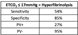
  • An ETCO2 value <17 mm Hg was determined to have a positive predictive value of 27% and a negative predictive value of 95%
  • The area under the receiver operating characteristic curve was 0.71, which was better than the blood pressure (0.58) and shock index (0.54)

The authors concluded that the ETCO2 was an accurate, objective, inexpensive, and noninvasive method of measuring the risk of hyperfibrinolysis that could guide the use of agents such as TXA.

Bottom line: A lot is going on in this abstract. The central concept is that it is trying to identify a surrogate for TEG-identified fibrinolysis available in the field. It compares ETCO2 with two other semi-objective indicators, blood pressure and shock index (pulse divided by blood pressure).

The biggest issue is that the number of patients with fibrinolysis was very small, only 13. Statistical comparisons of variables between the two groups are difficult because the number of HF patients in several subgroups was only 4 or 5. 

The sensitivity, specificity, and positive/negative predictive values are so-so. If the ETCO2 is above the 17mm Hg threshold, the likelihood of patients not having HF is good at 95%. But if it is below, the likelihood that they actually have HF is only 27% (true positive rate).
The area under the curve calculations is also not very impressive. Yes, an AUC of 0.71 is better than 0.54-0.58, but it is still not great.

One always has to be careful finding surrogates (ETCO2) for things you really want to measure (TEG LY30 > 3%). Many potential confounders can limit their usefulness. And this case is no different, which should be apparent from the numbers. Perhaps the data would be better if a much larger group of patients were studied. Unfortunately, this will probably take close to 1,000 subjects and require a multicenter trial. 

This is interesting preliminary work. It’s definitely not enough to change practice now. But with more work, and more patients, who knows?

Reference: Prehospital ETCO2 predicts hyperfibrinolysis in injured patients: implications for early use of antifibrinolytics in trauma. EAST 2024 Podium paper 3.

Best of EAST 2024 #1: MAP And Spinal Cord Injury

The use of elevated mean arterial pressure (MAP) to help manage spinal cord injury has been a mainstay of treatment for years. The concept is similar to that used for management of severe traumatic brain injury. The theory is that there may be areas of the brain that are damaged, but not irretrievably so. Increasing MAP should improve perfusion and may protect areas in jeopardy from secondary injury.

As with so much in neurotrauma, few large and/or prospective studies exist. Although most centers have specific algorithms and MAP goals, optimal treatment still needs to be determined.

EAST sponsored a prospective, multicenter study to identify factors influencing neurologic outcomes after spinal cord injury. MAP augmentation was monitored, specifically its impact on the American Spinal Injury Association (ASIA) score between admission and discharge.

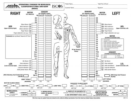
The ASIA Score is calculated by performing a very detailed exam consisting of myotomal motor function, a dermatomal sensory exam, and an anorectal exam. The exam takes quite some time to complete. The copy of the worksheet below should give you an idea of the level of detail:

The study was performed over 20 months, and 19 centers participated. They entered 222 patients, but only 164 had pre- and post-ASIA scores for comparison.

Here are the factoids:

  • Of the 164 patients studied, only 36 improved vs. 128 that showed no improvement by ASIA score
  • Demographics, hospital and ICU length of stay, and mortality were not significantly different between the groups
  • ISS was nearly identical (23 vs 25)
  • Three-quarters of injuries were to the cervical spine, about 10% to the lumbar spine, and the remainder to the thoracic spine. There was no correlation between injury location and recovery.
  • Presentation in the trauma bay (blood pressure, pulse, MAP, lactate, and Hgb) were the same in both groups
  • The MAP goal of >85 mm Hg was met about 75% of the time in both groups
  • Duration of MAP therapy was the same for the two groups, from 99-113 hours
  • There was a trend toward increased cardiac issues (atrial fibrillation, v-tach, elevated troponin) in the group with improved spinal cord recovery. This may be due to the medications used to increase MAP.

Bottom line: This is very interesting work and will make us question the utility of MAP therapy for spinal cord injury. However, this is not a cut-and-dried conclusion. Here are several things that come to mind:

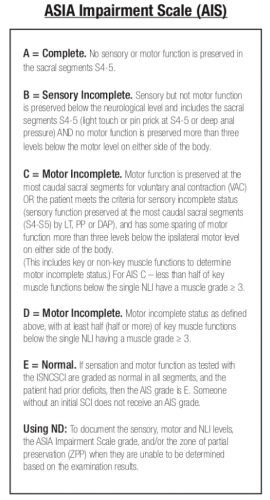
  • What was the definition of “improvement?” ASIA is a complicated scoring system with many steps in the evaluation. Usually, the results are condensed into an overall “ASIA Impairment Scale,” or AIS.
    The AIS is not very granular, meaning that each step in the scale represents a large difference in function. Could patients have had improvements that did not change the AIS score but were functionally significant for the patient? For example, an improvement from a C5 to a C6 level makes a big difference in daily activities.
  • Was the study large enough? It is difficult to accumulate a large series of spinal cord injury patients. Combining this point with the previous one, was the statistical power present even to detect a meaningful difference in the AIS?
  • Was MAP>85 torr maintained reliably and for long enough? Patients had MAP therapy for just over four days, and it was only maintained above the threshold about 75% of the time. We have good evidence in the brain injury literature that a single bout of hypotension in patients with severe TBI significantly increases mortality. Could it be that maintaining increased spinal cord perfusion is equally important? Could a single low MAP cause damage? It would be interesting to see if patients who had very consistent MAP therapy, say greater than 90% or 95% of the time, had any difference in outcomes. Unfortunately, I suspect that the numbers would be far too low to prove anything.

This abstract brings up some interesting questions. However, I would not consider throwing out the use of MAP goals based on it. We need more patients to study and be better at applying this treatment if we hope to uncover whether it really works.

Reference: Does mean arterial pressure augmentation improve neurological recovery of blunt spinal cord injuries: an EAST multicenter trial. EAST 2024 Podium paper #1.

Best Of AAST #5: Door-To-Prophylaxis Time

Today’s abstract continues the theme of VTE prophylaxis. The authors introduce another timing parameter in this one: the “door-to-prophylaxis” time. Just as it sounds, this is the time interval between admission to the ED and initiating chemo-prophylaxis. Just like some centers struggle with how long to wait to start it after a solid organ injury (see previous post here), many find it challenging to get it ordered in the first place.

The authors retrospectively reviewed their registry data over four years. They compared adult patients who arrived as a highest-level of activation and received blood during their resuscitation. They were divided into two groups: those with DVT or pulmonary embolism (VTE group) and those without (no VTE group). The door-to-prophylaxis time was defined as the time from hospital arrival to the first dose of medication.

Here are the factoids:

  • Just over 2,000 patients met inclusion criteria, with 106 experiencing VTE and 1,941 without it
  • VTE patients had higher ISS (29 vs. 24), higher lactate levels (4.6 vs. 3.9), and more post-ED blood transfusions (8 vs. 2)
  • There was no difference in the need for dose adjustment or missed doses between the groups
  • Door-to-prophylaxis time was significantly longer in the VTE group (35 vs. 25 hours)
  • When controlling for age, sex, ISS, lactate, and post-ED transfusions, each hour of delay increased the likelihood of VTE by 1.5%

The authors concluded that the door-to-prophylaxis time was significantly associated with increased incidence of VTE. They suggest that the door-to-prophylaxis time should be utilized as a performance improvement metric for this condition.

Bottom line: Unfortunately, we need a lot more information here. There was not enough room for details about the statistical analysis in the abstract, but they will be essential to know. And the authors remind us that this study shows association, not causation. 

Severe injury and blood transfusion are already known to be associated with a higher likelihood of VTE. The fact that the longer door-to-prophylaxis group had more frequent VTE may very well be due to their higher ISS and greater number of transfusions. Those events themselves may have led to the hesitation in starting a heparin.

Early prophylaxis is certainly a desirable goal in any trauma patient. But we need more than a new metric. We need more concrete information on the specific reasons for the delay and to prove that it is safe to give the drug early in patients who have those potential delaying factors.

Reference: “Door-to-prophylaxis” time as a novel quality improvement metric in preventing venous thromboembolism following traumatic injury. AAST 2023, Plenary paper #38.

Best Of AAST #4: Starting VTE Prophylaxis After Solid Organ Injury

Venous thromboembolic disease (VTE) continues to be a major issue in trauma patients. Most trauma centers have prophylaxis guidelines to try to reduce this problem. These guidelines typically recognize specific injuries that increase the risk of bleeding if anticoagulants are given. Typical ones include hemorrhagic injuries to the brain, pelvic and spine fractures, and solid organ injuries.

Typically, VTE prophylaxis starts immediately upon admission. But when these high-risk injuries are present, it is usually delayed for a period of time. Unfortunately, that period may be highly variable. Many centers have adopted 2-3 days to delay administration of low molecular weight heparin in patients with solid organ injury.

The AAST initiated a prospective multi-institutional trial comparing early (<48 hours after admission) and late (>48 hours) administration of prophylactic agents. Patients were older than 16 years, had any number of liver, spleen, or kidney injuries, and were initially treated nonoperatively. Patients who were transferred, died in the ED, were pregnant, had a bleeding disorder, or were taking anticoagulants or platelet inhibitors were excluded. A power analysis was performed, and more than the needed number of patients were enrolled.

Here are the factoids:

  • A total of 1173 patients were enrolled, and there were 589 liver injuries, 569 spleen injuries, and 289 kidney injuries
  • About 75% of patients (864) had early prophylaxis
  • Patients were younger (median 34 years), and two-thirds were male, with a median ISS of 22
  • Early VTE prophylaxis patients had significantly lower rates of VTE (3% vs. 7%)
  • There was no significant difference in failure of nonoperative management (5% early vs. 7% late)
  • The early prophylaxis group received fewer units of blood after prophylaxis started (17% vs. 23%)
  • Patients receiving VTE prophylaxis after 48 hours were 2.2x more likely to develop VTE

The authors concluded that early VTE chemoprophylaxis was associated with lower rates of VTE with no increase in complications. They recommended that it should become the standard of care for these patients.

Bottom line: Seeing such a well-designed and nicely executed study is refreshing. If the facts are borne out in the final manuscript review, this should become the standard of care for VTE prophylaxis in patients with solid organ injuries. 

I wish the authors would have stipulated that the chemoprophylaxis was required to be low molecular weight heparin. Unfortunately, there are still more than a few centers using unfractionated heparin. There could be a difference in efficacy and failure rates between the two. This could complicate the statistical analysis. Hopefully, the presenter will address this during the meeting.

I would also like to see a breakdown of when the early VTE prophylaxis actually started. Were they all close to 48 hours? Or were there enough at 24 hours to show this is also safe and effective?

It’s time for everyone to review their VTE prophylaxis guidelines. Get ready to make some major changes in your patients with solid organ injury!

Reference: When is it safe to start VTE prophylaxis after blunt solid organ injury? A prospective AAST multi-institutional trial. AAST 2023, Plenary paper #23.

Best Of EAST 2023 #12: VTE Prophylaxis In Severe TBI

Time for another abstract on venous thromboembolic disease (VTE) prophylaxis, but this time in patients with severe head injury. VTE is a significant problem for trauma patients. Those with a potential source of bleeding from their injuries cause us to hesitate and consider the timing of chemical prophylaxis closely. Do we really want to cause more bleeding?

This is particularly problematic with intracranial hemorrhage, as the treatment is major brain surgery. Over recent years, the literature has been leaning toward earlier prophylaxis as soon as the intracranial blood has stopped evolving.

The EAST Multicenter Trials Group performed a seven-year retrospective review at 24 Level I and II trauma centers to assess the safety and efficacy of VTE chemoprophylaxis.  They divided patients into three groups: no prophylaxis, early prophylaxis (within 24 hours), and late prophylaxis (after 24 hours).

The authors assessed two endpoints: VTE occurrence and expansion of intracranial hemorrhage (ICH). They used several regression models to check their hypotheses.

Here are the factoids:

  • A total of 2,659 patients met the inclusion criteria. This averages out to 15 eligible patients per month per center. This is probably reasonable when combining a few high-volume centers with more lower volume centers.
  • Compared to early prophylaxis, patients who received late prophylaxis were twice as likely to develop VTE, although this was not statistically significant (p = 0.059)
  • Compared to early prophylaxis, patients who received no prophylaxis were a third less likely to develop VTE, although this, too, was not statistically significant (p = 0.39
  • About 25% of patients who received either early or late prophylaxis suffered an extension of their ICH, but only 17% of the no-prophylaxis group did
  • The regression model showed that the no prophylaxis group was 36% less likely to develop ICH extension compared to either early or late prophylaxis groups.

The workgroup concluded that the development of VTE was not dependent on the timing of the start of prophylaxis. Furthermore, patients who did not receive any prophylaxis had significantly decreased odds of ICH extension. The group recommended larger randomized studies to extend this work.

Bottom line: Shocker! This multicenter study suggests that the no prophylaxis and early prophylaxis groups had fewer VTE events than the late group, although these results were not statistically significant. This means that there wasn’t an advantage to giving the shot.

And the other major conclusion was that both early and late prophylaxis was associated with a significantly higher incidence of ICH extension. 

Roll these together, and you will find that neither early nor late prophylaxis help prevent VTE, yet they are both associated with additional bleeding in and around the brain! 

Heresy! I am trying to figure out what to make of these results. Perhaps the retrospective nature of the study and the wildcards this introduces influenced the results. It could be a study power problem, except the numbers were approaching significance that was unfavorable for prophylaxis.

I will be very interested to hear how the authors explain these findings. And yes, a well-powered randomized study would be great, but I don’t think many institutional review boards will be keen on a no-treatment group given our current fear of VTE. So don’t count on any real answers soon.

Reference: EARLY VTE PROPHYLAXIS IN SEVERE TRAUMATIC BRAIN INJURY: A PROPENSITY SCORE WEIGHTED EAST MULTICENTER TRIAL. EAST 2023 Podium paper #38.