Tag Archives: abdomen

Gunshots And CT Scan Of The Abdomen

Abdominal gunshots and CT scanning are usually thought to be mutually exclusive. The usual algorithm generally means a prompt trip to the operating room. But as with many things in the management of trauma, there are always exceptions. The key is to understand when exactly one of those exceptions is warranted.

Exception 1: Did it really enter the abdomen? Gunshots have enough energy that they usually do get inside. However, freaky combinations of trajectory and body habitus do occur. There are three tests that must be passed in order to entertain the possibility that the bullet may not have made it inside your patient: physiology, anatomy, and physical exam. For physiology, the patient must be completely hemodynamically stable. Anatomically, the trajectory must make sense. If the known wounds and angles allow a tangential course make sense, then fine. But if there is a hole in the epigastrium and another next to the spine, you have to assume the bullet went straight through. Finally, the physical exam must be normal. No peritonitis. No generalized guarding. Focal tenderness only in the immediate area of any wounds. If all three of these criteria are passed, then a CT can be obtained to demonstrate the trajectory.

Exception 2: Did it enter an unimportant area of the abdomen? Well, there’s really only one of these, and that’s the area involving the right lobe of the liver and extending posteriorly and lateral to it. If the bullet hole(s) involve only this area, and the three tests above are passed, CT may confirm an injury that can be observed. However, there should only be a minimal amount of free fluid, and no soft tissue changes of any kind adjacent to bowel.

Exception 3: A prompt trauma lap was performed, but you think you need more information afterwards. This is rare. The usual belief is that the eyes of the surgeon provide the gold standard evaluation during a trauma lap. For most low velocity injuries with an easily understood trajectory, this is probably true. However, high velocity injuries, those involving multiple projectiles, or complicated trajectories (side to side) can be challenging for even the most experienced surgeon. Some areas (think retroperitoneum or deep in the pelvis) are tough to visualize completely, especially when there’s blood everywhere. These are also the cases most likely to require damage control surgery, so once the patient has been temporarily closed, warmed and resuscitated, a quick trip to CT may be helful in revealing unexpected shrapnel, unsuspected injuries, or other issues that may change your management. Even a completely unsurprising scan can provide a higher sense of security.

Bottom line: CT of the abdomen and gunshots to that area may actually coexist in some special cases. Make sure the physiology, anatomy and physical exam criteria are passed first. I also make a point of announcing to all trainees that taking these patients to CT is not the norm, and carefully explain the rationale. Finally, apply the concept of the null hypothesis to this situation. Your null hypothesis should state that your patient does not need a CT after gunshot to the abdomen, and you have to work to prove otherwise!

What The Heck? Bicycle Trauma Answer

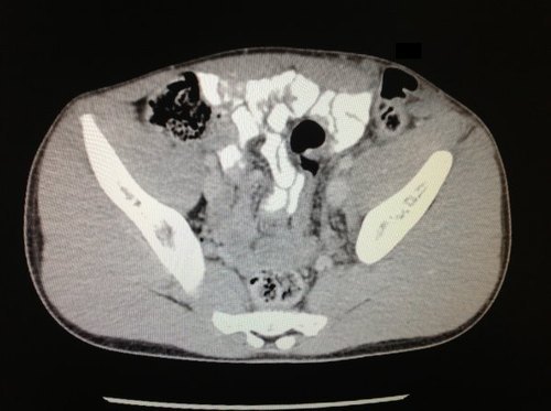
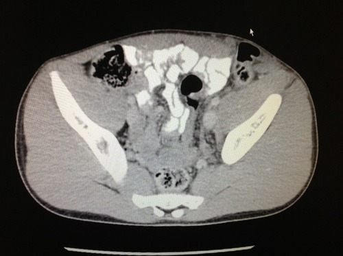
So a young male jammed a handlebar into his abdomen, and a CT image demonstrating his problem was shown. But what did it actually show?

By now, you probably realize that clinical information is key. On exam, he had an obvious bulge in his left lower quadrant, more obvious with straining. Looking at the CT (now with a nice arrow), there is a problem over the left side of the abdomen. 

image

This child has so little fat, that it’s difficult to see the problem. If you track the thin layer of fat across the abdomen to the right side of the image, you’ll see that it disappears over the bowel gas. This represents a complete tear through all fascial layers, not just a Spigelian hernia as some readers guessed.

Management consisted of primary repair of the defect. An uneventful recovery can be expected. Unless more bicycle tricks are anticipated.

Reference: Traumatic handlebar hernia: a rare abdominal wall hernia. J Ped Surg 39(10):e20-e22, 2004.

What The Heck? Bicycle Trauma Part 2

Yesterday’s puzzle involved a young male who drove a handlebar into his abdomen. Little additional information was given, other than one slice of his abdominal CT scan. So what’s the problem?

The textbooks always associate handlebars with pancreatic and duodenal injuries, and these should always be looked for. However, the scan slice in this case was taken lower, within the pelvis. Too low to show you either of those organs.

As I’ve said before, be systematic when reading xray images. We automatically focus on the viscera and bones. Look at those areas, make sure you can identify each structure that you see, and look for any anomalies. 

But don’t forget the soft tissue! In this case, the child doesn’t have much. Take a closer look at the same slice and see if you can figure it out by tomorrow.

Sonography In Place of CT For Pediatric Abdominal Trauma

Pediatric blunt abdominal trauma is not common, but if present it has the potential to cause significant morbidity or mortality. Evaluation of the abdomen at the trauma center is crucial, and most trauma professionals are aware of the trade-offs in the use of CT scan in children (radiation exposure, need for sedation).

Ultrasound is widely available and allows for imaging of most areas of concern in the abdomen. Could sonography be used in place of CT in specific cases? Pediatric surgeons in Germany (who have been using ultrasound far longer than the US has) published a paper last year looking at their experience with children who were diagnosed with an intra-abdominal organ injury after blunt trauma. Their 7 year experience only produced 35 such children, and they were evaluated with examination and one or more serial FAST ultrasound exams. Equivocal results were scanned with CT.

They found that ultrasound was effective in diagnosing abdominal injury 97% of the time. Although 11 of the 35 children had subsequent CT scanning, it only changed management in one case

Bottom line: Obviously, this is a very small retrospective series, but it is provocative. The German pediatric surgeons go above and beyond the typical FAST exam in the US, using it for diagnostic purposes as well. Could a complete diagnostic ultrasound take the place of CT in select children in the US? Probably so, as they are very sensitive in detecting free fluid and solid organ injury. But what about blunt intestinal injury? I’ll review that tomorrow and sum up my thoughts on a possible algorithm.

Related posts:

Reference: Is sonography reliable for the diagnosis of pediatric blunt abdominal trauma? J Pediatric Surg 45(5):912-915, 2010.

Trauma 20 Years Ago: Seatbelt Injuries

Seatbelt use has increased from 58% in 1994 to a high of 85% last year. We know that seatbelt use saves lives, but trauma professionals are also aware that they can create their own injuries as well. This is a positive trade-off, because belt use prevents injuries that are difficult to treat (e.g. severe brain injury) and produces a higher number of intra-abdominal injuries that are easy to treat.

The spectrum of injuries attributed to seat belt use was finally appreciated in a journal article published 20 years ago this month. The authors wanted to catalog the various injuries seen in belted and unbelted motor vehicle occupants. They reviewed data from the North Carolina Trauma Registry, one of the most sophisticated state registries at the time. Although there were over 21,000 records in the database, only 3,901 involved motor vehicle crashes and had complete data on seatbelt use.

This study found the following:

  • Mortality was higher in those not wearing their seat belts (7% vs 3.2%)
  • Unbelted had a much higher incidence of severe head injury (50% vs 33%)
  • Overall incidence of any abdominal injury was the same for both (14%)
  • GI tract injuries were more common in the belted group (3.4% vs 1.8%)
  • Solid organ injury was the same

Bottom line: This study sparked the recognition that seatbelts reduce severe head injury but increase the incidence of some hollow viscus injuries. About 514 severe head injuries were prevented in exchange for 21 additional abdominal injuries that were generally easily repaired. Good tradeoff!

Related posts: 

Reference: The spectrum of abdominal injuries associates with the use of seat belts. J Trauma 31(6):821-826, 1991.