Category Archives: Thorax

Detecting Rib Fractures In The Elderly

It’s well known that our elders do less well than younger folks after injury. The number of complications is higher, there tends to be more loss of independence during recovery, and mortality is increased. This is not only true of high energy trauma like car crashes, but also much lower energy events such as a fall from standing.

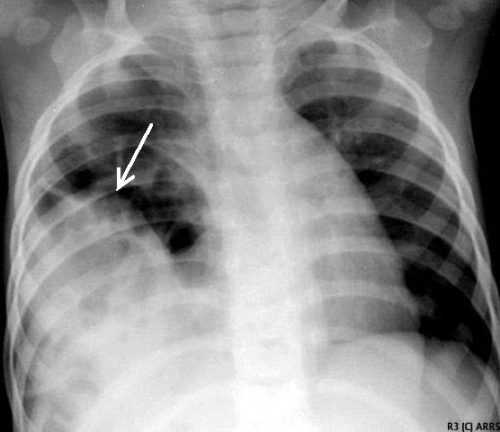
Rib fractures are common after falls in the elderly and contribute to significant morbidity if not treated adequately. Traditionally, they are identified through a combination of physical exam and chest x-ray. Unfortunately, only half of rib fractures are visible on x-ray. It falls to the physical exam to detect the rest.

A group at Beth Israel Hospital in Boston explored the utility of using chest CT in an attempt to determine if this would result in more appropriate and cost-efficient care in the elderly. They performed a retrospective study of 3 years of their own data on patients aged 65 or more presenting after a mechanical fall and receiving a rib fracture diagnosis. Imaging was ordered at the discretion of the physician. A total of 330 patients were elderly, fell, and had both chest x-ray and chest CT obtained. This was a very elderly group, with a mean age of 84 years!

Here are the factoids:

  • Rib fractures were seen on chest x-ray in 40 patients (12%) and on CT in an additional 56 ; 234 patients had no fractures on either
  • When fractures were seen on both studies, CT identified a median of 2 more fractures than chest x-ray
  • Patients with fractures not seen on chest x-ray were admitted significantly more often than those without fractures (91% vs 78%)
  • Mortality, admission to ICU, ICU length of stay, and hospital length of stay were not different if fractures were seen only on CT
  • CT scan identified new issues or clarified diagnoses suggested by chest x-ray in 14 cases, including one malignancy
  • Rib detail images were obtained in 13 patients and proved to be better than chest x-ray, but not quite as good as CT scan

Conclusion: use of CT for rib fracture diagnosis resulted in a few more admissions, but no change in hospital resource utilization, complications, or mortality.

Bottom line: Hmm…, read the paper closely. The authors conclude that more patients with CT-only identified rib fractures are admitted. But compared to what? Unfortunately, patients without rib fractures on CT. What about comparing to patients who had fractures seen on chest x-ray too? If that number is the same, then of what additional use is CT? Identifying a few incidentalomas?

Given that there is no change in the usual outcome measures listed here, it doesn’t seem like there is any additional benefit to adding CT. And I can see a lot of downsides: cost, radiation, and possible exposure to IV contrast. In my mind, there is still nothing that beats a good physical exam and a chest x-ray. Skip the CT scan. And don’t even think about ordering rib detail images! That’s so 1990s. And even if no rib fractures are seen on imaging, physical exam is the prime determinant for admitting your patient for aggressive pain management and pulmonary toilet.

Reference: Chest CT imaging utility for radiographically occult rib fractures in elderly fall-injured patients. J Trauma 86(5):838-843, 2019.

Delayed Presentation Of Right Diaphragm Injury

Diaphragm injury from blunt trauma is uncommon, occurring in only a few percent of patients after high energy mechanisms. They usually occur on the left side, and are more frequently seen after t-bone type car crashes and in pedestrians struck by a car.

Blunt diaphragm injury on the right side is very unusual. Even so, it is more easily detected due to obvious displacement of the liver that can be seen on chest x-ray. Blunt injuries on the right side usually result in a large rent in the central tendon, or detachment of the diaphragm from the chest wall. This allows the liver to herniate into the chest, and the chest x-ray finding is not subtle.

This image shows an acute herniation of the liver through the diaphragm. Due to the size of the liver, only part of it can typically fit through the rent. Radiologists call this the “cottage loaf” sign. Why? Here’s the bakery item it is named after. Get it now?

Thankfully, most of these injuries are identified in the acute setting. They must be addressed surgically because, if left untreated, more and more of the liver will slowly move into the chest resulting in respiratory problems in the long run.

Acute management usually consists of laparotomy to address both the diaphragm tear and any other associated intra-abdominal injuries. The liver should be reduced by sliding a hand next to it laterally into the chest cavity and pushing the dome downwards. The right triangular ligaments should be taken down (if they are not already destroyed) to mobilize the organ better so the diaphragm laceration can be closed. This is typically accomplished with some type of large (size 0) permanent suture. A chest tube will be needed to evacuate the iatrogenic pneumothorax created by opening the abdomen.

Chronic right diaphragm injuries are a different animal entirely. There is no longer any need to evaluate for intra-abdominal injury, so the procedure is usually performed through the chest. For smaller injuries, thoracoscopic procedures have been described that push the liver downwards and then either suture the diaphragm primarily or (more likely) incorporate a piece of mesh.

Larger injury requires conversion to an open procedure so more muscle power can be used to push the liver downwards to facilitate the repair. However, do not underestimate the adhesions that will be present between diaphragm and liver (and possibly the lung) in long-standing injuries. It may take some time to dissect them away. Rarely, a laparotomy (or laparoscopy) may be needed to assist for very large and complex injuries.

References:

  • Management of Delayed Presentation of a Right-Side Traumatic
    Diaphragmatic Rupture. World J Surg 36:260-265, 2012.
  • Delayed Discovery of Diaphragmatic Injury After Blunt Trauma:
    Report of Three Cases. Surg Today 35:407-410, 2005.

What Is A Wide Mediastinum Anyway?

Trauma professionals are always on the lookout for injuries that can kill you. Thoracic aortic injury from blunt trauma is one of those injuries. Thankfully, it is uncommon, but it can certainly be deadly.

One of the screening tests used to detect aortic injury is the old-fashioned chest xray. This test is said to be about 50% sensitive, with a negative predictive value of about 80%. However, the sensitivity is probably decreasing and the negative predictive value increasing due to the rapidly increasing number of obese patients that we see.

A wide mediastinum is defined as being > 8cm in width. In this day and age of digital imaging, you will need to use the measurement tool on your workstation to figure this out.

Unfortunately, it seems like most chest xrays show wide mediastinum these days. What are the most common causes for this?

  • Technique. The standard xray technique used to reduce magnification of the anterior mediastinum (where the aortic arch lives) is a tube distance of 72 inches from the patient, shot back to front. We can’t do this for trauma patients because we can’t stand them up and are reluctant to prone them. The standard trauma room technique is 36 inches from the patient shot front to back. This serves to magnify the mediastinal image and make it look wide.
  • Obesity. The more fat in the mediastinum, the wider it looks. The more fat on the back, the further the mediastinum is from the xray plate and the greater the magnification.
  • Other mediastinal blood. Major blunt trauma to the chest can cause bleeding from small veins in the mediastinum, making it look wide.
  • Thymus. Only in kids, though.
  • Aortic injury. Last but not least. Only a few percent of people with wide mediastinum will actually have the injury.

If you encounter a wide mediastinum on chest xray in a patient with a significant mechanism for aortic injury, then they should be screened using helical CT.

How To: Needle Decompression Of The Chest

Here’s a quick, 3 ½ minute video for physicians and paramedics on how to decompress the chest when you suspect a tension pneumothorax.

The ATLS course now adds a consideration to use an alternative site. That location is the 5th intercostal space around the mid-axillary line. This has come about because shorter needles may not reach the pleural space when inserted under the clavicle in larger patients. The new spot is the typical location for placement of the inevitable chest tube that has to be inserted after needle decompression.

If you’ve got a few tips or tricks that you’d like to share on this procedure, please comment on the YouTube video.

YouTube player

Best Of EAST 2020 #3: Rib Fixation In The Elderly

Elderly falls have reached epidemic proportions. Although the most common injury from these falls is rapidly become head injury with or without intracranial blood, rib fractures are a close second. Treatment of rib fractures usually involves multiple interventions such as pulmonary toilet, multimodal pain management, and therapies to enhance mobility. And in some cases, operative fixation is entertained.

Rib fracture fixation has typically been used in patients who are dependent on a ventilator due to their fractures, or have significantly displaced or very painful fractures. There is little data on the impact of using rib plating in elderly patients. The group at New York Presbyterian Hospital in Queens NY analyzed one year of TQIP data to assess the impact of this technique in trauma patients older than 65.

They reviewed the data, looking at mortality, intensive care unit and hospital lengths of stay, tracheostomy, and pneumonia rates. They matched patients who had rib fixation with similar patients who did not. They then sliced and diced the data to see if there were differences in these outcomes with early vs late (> 48hrs) repair.

Here are the factoids:

  • The authors obtained data on almost 14,000 patients meeting study criteria, but of those only 278 underwent rib fixation and 220 were eligible for matching
  • Overall, patients who underwent fixation tended to have higher rates of flail chest, earlier intubation, higher injury severity score, and increased intensive care unit admission rates
  • Mortality for all patients who underwent fixation was significantly lower than those who did not (4% vs 10%)
  • The early fixation group had significantly fewer ventilator associated penumonias, shorter ICU length of stay (6 vs 10 days) and shorter hospital length of stay (9 vs 15)
  • There were no differences in mortality or ventilator days

The author’s conclusions matched the bullet items above.

My comment: This is one of those papers that demonstrates something that we should have already recognized. I wish I had thought of it! It points us toward considering this procedure in our elderly rib fracture patients. Even though patients undergoing fixation were sicker and had more serious injuries, their survival rate was significantly higher.

However, it also leaves us with more work to do. It is a database study, so it’s not possible to go back and find additional information on the study subjects. Knowing selection criteria and operative details would be very helpful. And the overall numbers are low, so more benefits may come to light if we had the statistical power to focus on mortality and ventilator days.

Here are my questions for the authors and presenter:

  1. Have you considered using a larger dataset to get additional information? The mortality and ventilator days in the early vs late subsets were not statistically significant. This might be due to the lack of statistical power from the small number of patients.
  2. Can you speculate on the financial impact of using expanding the use of rib fixation in the elderly? The clinical impact is clear. It looks like the cost savings to the hospital from the reduced ICU and hospital length of stay alone would far offset the cost of performing the procedure, especially if done early.
  3. What selection criteria should be used for choosing the right patients for the procedure? This is probably outside the scope of the study, but it would be interesting to hear you speculate.

This is an important paper and I really look forward to hearing the details!

Reference: Rib fixation in geriatric trauma: mortality benefits for the most vulnerable patients. EAST Annual Assembly abstract #3, 2020.