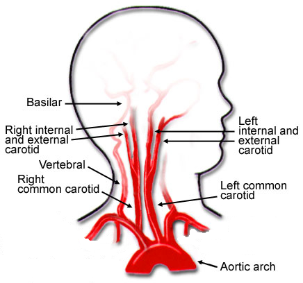
Category Archives: Thorax

Delayed Hemothorax In Older Adults: Real Or Not?

I came across an interesting paper in the Journal of Trauma & Acute Care Surgery Open recently. I always read these articles a bit more critically, though, because the peer review process just doesn’t feel quite the same to me as the more traditional journal process. But maybe it’s just me.

In this paper, the authors decided to look at the incidence of delayed hemothorax because “emerging evidence suggests HTX in older adults with rib fractures may experience subtle hemothoraces that progress in a delayed fashion over several days.” They cite two references to back up this rationale.

They retrospectively reviewed records from two busy US Level I trauma centers for adults age 50 or older who were diagnosed with delayed hemothorax (dHTX). Delayed was defined as 48 hours or more after initial chest CT showed either a minimal or trace HTX. The authors went on to analyze the characteristics and demographics of the patients involved.

Here are the factoids:

  • A total of 14 older adults experienced dHTX after rib fractures, an overall incidence of 1.3% (!)
  • About half were diagnosed during the initial hospitalization for the fractures
  • All patients had multiple fractures, with an average of 6 consecutive ones; four had a flail chest
  • One third progressed from a trace HTX, two thirds had a completely negative initial chest CT
  • Only one third were taking anticoagulants or anti-platelet agents
  • Patients with multiple fractures, posteriorly located, and displaced were most likely to develop dHTX

The authors concluded that “delayed progression and delayed development of HTX among older adults with rib fractures require wider recognition.”

Bottom line: Really? First, I looked at the papers cited by the authors as the rationale for doing this study. They each found dHTX in about 10% of patients, but their definition was very broad: any fluid visible on upright chest x-ray. Furthermore, the patients were not really “older” either. Average age was around 50. 

So I’m not sure yet whether this is a problem, especially with the low incidence of 1.3%. This study doesn’t come right out and state how many patients they reviewed to find their 14, but it can be calculated to be 14 / 1.3% = 1,177. This incidence is only one tenth of that found in the two studies cited. Seems relatively uncommon, and half were discovered while the patients were still in the hospital. Thus only 0.65% sought readmission for chest discomfort or difficulty breathing.

This study required chest CT for rib fracture diagnosis. Is all that radiation (and possibly contrast) really necessary? And did these patients get another chest CT to delineate the pathology? More radiation?

Overall, this paper was not very helpful to me. Yes, I have seen patients come back days or weeks later with a hemothorax that was not seen during their first visit. It’s just that this study raises many more questions that should have been easily answered in the discussion. But they weren’t.

Given that only about a half of a percent of rib fracture patients develop delayed hemothorax after discharge, it is probably prudent to provide information to the patient recommending they see their practitioner if they develop any symptoms days or weeks later.  And a simple chest x-ray should do.

Reference: Complication to consider: delayed traumatic hemothorax in older adults. Trauma Surgery & Acute Care Open 2021;6:e000626. doi: 10.1136/tsaco-2020-000626.

Print Friendly, PDF & Email

Detecting Rib Fractures In The Elderly

It’s well known that our elders do less well than younger folks after injury. The number of complications is higher, there tends to be more loss of independence during recovery, and mortality is increased. This is not only true of high energy trauma like car crashes, but also much lower energy events such as a fall from standing.

Rib fractures are common after falls in the elderly and contribute to significant morbidity if not treated adequately. Traditionally, they are identified through a combination of physical exam and chest x-ray. Unfortunately, only half of rib fractures are visible on x-ray. It falls to the physical exam to detect the rest.

A group at Beth Israel Hospital in Boston explored the utility of using chest CT in an attempt to determine if this would result in more appropriate and cost-efficient care in the elderly. They performed a retrospective study of 3 years of their own data on patients aged 65 or more presenting after a mechanical fall and receiving a rib fracture diagnosis. Imaging was ordered at the discretion of the physician. A total of 330 patients were elderly, fell, and had both chest x-ray and chest CT obtained. This was a very elderly group, with a mean age of 84 years!

Here are the factoids:

  • Rib fractures were seen on chest x-ray in 40 patients (12%) and on CT in an additional 56 ; 234 patients had no fractures on either
  • When fractures were seen on both studies, CT identified a median of 2 more fractures than chest x-ray
  • Patients with fractures not seen on chest x-ray were admitted significantly more often than those without fractures (91% vs 78%)
  • Mortality, admission to ICU, ICU length of stay, and hospital length of stay were not different if fractures were seen only on CT
  • CT scan identified new issues or clarified diagnoses suggested by chest x-ray in 14 cases, including one malignancy
  • Rib detail images were obtained in 13 patients and proved to be better than chest x-ray, but not quite as good as CT scan

Conclusion: use of CT for rib fracture diagnosis resulted in a few more admissions, but no change in hospital resource utilization, complications, or mortality.

Bottom line: Hmm…, read the paper closely. The authors conclude that more patients with CT-only identified rib fractures are admitted. But compared to what? Unfortunately, patients without rib fractures on CT. What about comparing to patients who had fractures seen on chest x-ray too? If that number is the same, then of what additional use is CT? Identifying a few incidentalomas?

Given that there is no change in the usual outcome measures listed here, it doesn’t seem like there is any additional benefit to adding CT. And I can see a lot of downsides: cost, radiation, and possible exposure to IV contrast. In my mind, there is still nothing that beats a good physical exam and a chest x-ray. Skip the CT scan. And don’t even think about ordering rib detail images! That’s so 1990s. And even if no rib fractures are seen on imaging, physical exam is the prime determinant for admitting your patient for aggressive pain management and pulmonary toilet.

Reference: Chest CT imaging utility for radiographically occult rib fractures in elderly fall-injured patients. J Trauma 86(5):838-843, 2019.

Print Friendly, PDF & Email

Delayed Presentation Of Right Diaphragm Injury

Diaphragm injury from blunt trauma is uncommon, occurring in only a few percent of patients after high energy mechanisms. They usually occur on the left side, and are more frequently seen after t-bone type car crashes and in pedestrians struck by a car.

Blunt diaphragm injury on the right side is very unusual. Even so, it is more easily detected due to obvious displacement of the liver that can be seen on chest x-ray. Blunt injuries on the right side usually result in a large rent in the central tendon, or detachment of the diaphragm from the chest wall. This allows the liver to herniate into the chest, and the chest x-ray finding is not subtle.

This image shows an acute herniation of the liver through the diaphragm. Due to the size of the liver, only part of it can typically fit through the rent. Radiologists call this the “cottage loaf” sign. Why? Here’s the bakery item it is named after. Get it now?

Thankfully, most of these injuries are identified in the acute setting. They must be addressed surgically because, if left untreated, more and more of the liver will slowly move into the chest resulting in respiratory problems in the long run.

Acute management usually consists of laparotomy to address both the diaphragm tear and any other associated intra-abdominal injuries. The liver should be reduced by sliding a hand next to it laterally into the chest cavity and pushing the dome downwards. The right triangular ligaments should be taken down (if they are not already destroyed) to mobilize the organ better so the diaphragm laceration can be closed. This is typically accomplished with some type of large (size 0) permanent suture. A chest tube will be needed to evacuate the iatrogenic pneumothorax created by opening the abdomen.

Chronic right diaphragm injuries are a different animal entirely. There is no longer any need to evaluate for intra-abdominal injury, so the procedure is usually performed through the chest. For smaller injuries, thoracoscopic procedures have been described that push the liver downwards and then either suture the diaphragm primarily or (more likely) incorporate a piece of mesh.

Larger injury requires conversion to an open procedure so more muscle power can be used to push the liver downwards to facilitate the repair. However, do not underestimate the adhesions that will be present between diaphragm and liver (and possibly the lung) in long-standing injuries. It may take some time to dissect them away. Rarely, a laparotomy (or laparoscopy) may be needed to assist for very large and complex injuries.

References:

  • Management of Delayed Presentation of a Right-Side Traumatic
    Diaphragmatic Rupture. World J Surg 36:260-265, 2012.
  • Delayed Discovery of Diaphragmatic Injury After Blunt Trauma:
    Report of Three Cases. Surg Today 35:407-410, 2005.
Print Friendly, PDF & Email

What Is A Wide Mediastinum Anyway?

Trauma professionals are always on the lookout for injuries that can kill you. Thoracic aortic injury from blunt trauma is one of those injuries. Thankfully, it is uncommon, but it can certainly be deadly.

One of the screening tests used to detect aortic injury is the old-fashioned chest xray. This test is said to be about 50% sensitive, with a negative predictive value of about 80%. However, the sensitivity is probably decreasing and the negative predictive value increasing due to the rapidly increasing number of obese patients that we see.

A wide mediastinum is defined as being > 8cm in width. In this day and age of digital imaging, you will need to use the measurement tool on your workstation to figure this out.

Unfortunately, it seems like most chest xrays show wide mediastinum these days. What are the most common causes for this?

  • Technique. The standard xray technique used to reduce magnification of the anterior mediastinum (where the aortic arch lives) is a tube distance of 72 inches from the patient, shot back to front. We can’t do this for trauma patients because we can’t stand them up and are reluctant to prone them. The standard trauma room technique is 36 inches from the patient shot front to back. This serves to magnify the mediastinal image and make it look wide.
  • Obesity. The more fat in the mediastinum, the wider it looks. The more fat on the back, the further the mediastinum is from the xray plate and the greater the magnification.
  • Other mediastinal blood. Major blunt trauma to the chest can cause bleeding from small veins in the mediastinum, making it look wide.
  • Thymus. Only in kids, though.
  • Aortic injury. Last but not least. Only a few percent of people with wide mediastinum will actually have the injury.

If you encounter a wide mediastinum on chest xray in a patient with a significant mechanism for aortic injury, then they should be screened using helical CT.

Print Friendly, PDF & Email

How To: Needle Decompression Of The Chest

Here’s a quick, 3 ½ minute video for physicians and paramedics on how to decompress the chest when you suspect a tension pneumothorax.

The ATLS course now adds a consideration to use an alternative site. That location is the 5th intercostal space around the mid-axillary line. This has come about because shorter needles may not reach the pleural space when inserted under the clavicle in larger patients. The new spot is the typical location for placement of the inevitable chest tube that has to be inserted after needle decompression.

If you’ve got a few tips or tricks that you’d like to share on this procedure, please comment on the YouTube video.

YouTube player
Print Friendly, PDF & Email