Trauma professionals are always on the lookout for injuries that can kill you. Thoracic aortic injury from blunt trauma is one of those injuries. Thankfully, it is uncommon, but it can certainly be deadly.
One of the screening tests used to detect aortic injury is the old-fashioned chest xray. This test is said to be about 50% sensitive, with a negative predictive value of about 80%. However, the sensitivity is probably decreasing and the negative predictive value increasing due to the rapidly increasing number of obese patients that we see.
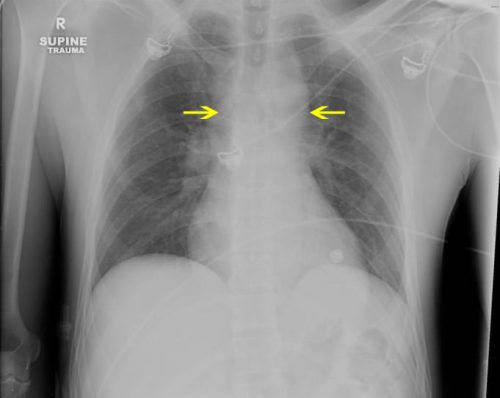
A wide mediastinum is defined as being > 8cm in width. In this day and age of digital imaging, you will need to use the measurement tool on your workstation to figure this out.
Unfortunately, it seems like most chest xrays show wide mediastinum these days. What are the most common causes for this?
- Technique. The standard xray technique used to reduce magnification of the anterior mediastinum (where the aortic arch lives) is a tube distance of 72 inches from the patient, shot back to front. We can’t do this for trauma patients because we can’t stand them up and are reluctant to prone them. The standard trauma room technique is 36 inches from the patient shot front to back. This serves to magnify the mediastinal image and make it look wide.
- Obesity. The more fat in the mediastinum, the wider it looks. The more fat on the back, the further the mediastinum is from the xray plate and the greater the magnification.
- Other mediastinal blood. Major blunt trauma to the chest can cause bleeding from small veins in the mediastinum, making it look wide.
- Thymus. Only in kids, though.
- Aortic injury. Last but not least. Only a few percent of people with wide mediastinum will actually have the injury.
If you encounter a wide mediastinum on chest xray in a patient with a significant mechanism for aortic injury, then they should be screened using helical CT.
