Category Archives: Thorax

Best Of AAST 2021: Chest Tube Based On Pneumothorax Size

How big is too big? That has been the question for a long time as it applies to pneumothorax and chest tubes. For many, it is a math problem that takes into account the appearance on chest x-ray, the physiology of the patient, and their ability to tolerate the pneumothorax based on any pre-existing medical conditions.

The group at Froedtert in Milwaukee has been trying to make this decision a bit more objective. They introduced the concept of CT based size measurement using a 35mm threshold at this very meeting three years ago. Read my review here. My criticisms at the time centered around the need to get a CT scan for diagnosis and their subjective definition of a failure requiring chest tube insertion. The abstract never did make it to publication.

The authors are back now with a follow-on study. This time, they made a rule that any pneumothorax less than 35mm from the chest wall would be observed without tube placement. The performed a retrospective review of their experience and divided it into two time periods: 2015-2016, before the new rule, and 2018-2019, after the new rule. They excluded any chest tubes inserted before the scan was performed, those that included a sizable hemothorax, and patients placed on a ventilator or who died.

Here are the factoids:

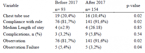
  • There were 93 patients in the early period and 154 in the later period
  • Chest tube use significantly declined from 20% to 10% between the two periods
  • Compliance with the rule significantly increased from 82% to 92%
  • There was no difference in length of stay, complications, or death
  • Observation failure was marginally less in the later period, and statistical significance depends on what method you use to calculate it
  • Patients in the later group were 2x more likely to be observed (by regression analysis)

The authors concluded that the 35mm rule resulted in a two-fold increase in observation and decreased the number of unnecessary CT scans.

Bottom line: I still have a few issues with this series of abstracts. First, decision to insert a chest tube requires a CT scan in a patient with a pneumothorax. This seems like extra radiation for patients who may not otherwise fit any of the usual blunt imaging criteria. And, like their 2018 abstract, there is no objective criteria for failure requiring tube insertion. So the number of insertions can potentially be quite subjective based on the whims of the individual surgeon.

What this abstract really shows is that compliance with the new rule increased, and there were no obvious complications from its use. The other numbers (chest tube insertions, observation failure) are just too subjective to learn much from.

Here are my questions for the presenter and authors:

  • Why was there such a large increase in the number of subjects for two identical-length time periods? Both were two years long, yet there were two-thirds more patients in the later period. Did your trauma center volumes go up that much? If not, could this represent some sort of selection bias that might change your numbers?
  • You concluded that your new rule decreased the number of “unnecessary” CT scans? How so? It looks like you are using more of them!
  • Do you routinely get a chest CT on all your patients with pneumothorax? Seems like a lot of radiation just to decide whether or not to put a tube in.
  • How do you manage a pneumothorax found on chest x-ray? Must they get a CT? Or are you willing to watch them and follow with serial x-rays?
  • How do you decide to take out the chest tube? Hopefully not another scan!

There should be some very interesting discussion of this abstract!

Reference: THE 35-MM RULE TO GUIDE PNEUMOTHORAX MANAGEMENT: INCREASES APPROPRIATE OBSERVATION AND DECREASES UNNECESARY CHEST TUBES. AAST 2021, Oral abstract #56.

Best Of AAST 2021: Liposomal Bupivacaine For Rib Fractures

The mainstays of rib fracture management are pain control and pulmonary toilet. The pain part of the equation can be managed in many ways, using topical, oral, IV, and injectable medications.

Rib blocks have been a mainstay for achieving some degree of local pain control. Classically, xylocaine was injected in the area around the costal nerve at or proximal to the fracture site. Then we found that if we combined the anesthetic agent with epinephrine, we could prolong the effect. New, longer-acting agents came around, and we could achieve a longer duration of action.

Then there is the new kid on the block: liposomal bupivacaine, also known as Exparel in the US.  The manufacturer was able to take molecules of bupivacaine and encapsulate them in a lipid membrane. When injected, these little liposomes slowly release their cargo, with a more prolonged anesthetic effect. Allegedly.

Sounds great! But does it work? The group at University of Cincinnati designed a prospective, double-blinded, randomized placebo control study of liposomal bupivacaine vs saline injection for pain control in up to six rib fractures. Subjects had significant injury as measured by their inability to achieve at least 50% of the desired inspiratory capacity. The authors monitored a number of respiratory parameters, as well as the pain score.

Here are the factoids:

  • Two cohorts of 50 patients were recruited, one received liposomal bupivacaine in up to six rib fractures, and the other received saline injections
  • The bupivacaine group achieved higher incentive spirometry volumes over the first two days, by about 200 cc
  • There was no change in daily pain scores in either group
  • Both groups showed a similar decrease in opioid use over time
  • Hospital and ICU lengths of stay were the same, and there were no complications or adverse events

Bottom line: Hmm. What’s going on here? There is a moderate amount of literature out there that does indicate a positive effect from liposomal bupivacaine in other conditions. But there are also some blinded, randomized studies that fail as well. So there are three possibilities:

  1. Liposomal bupivacaine isn’t a panacea, and works better in some situations than others
  2. This study failed to show a real difference for some reason
  3. A combination of both

This is a relatively small study, and the authors were not able to share their power analysis. They did not state if the spirometry volumes were significantly different, although I’m not sure 200 cc is clinically relevant. Maybe. But pain scores remained similar and opioid use declined as expected in both. 

These kinds of studies can be important. The difference in cost between injecting liposomal bupivacaine ($19 / ml) vs regular bupivacaine (10 cents / ml) vs saline/nothing (free) is striking. The premium price for the liposomal form needs to have a clear benefit or a cheaper product should be used.

Here are my questions for the presenter and authors:

  • Was your study big enough to show a result? Show us your power analysis.
  • How significant was the incentive spirometry result. Was the difference clinically noticeable?
  • What is your takeaway for this study? Your conclusion parrots the results. What will you do differently now, if anything?

Reference: INTERCOSTAL LIPOSOMAL BUPIVACAINE INJECTION FOR
RIB FRACTURES. AAST 2021, Oral abstract #20.

How Common Is BCVI, Really?

Blunt carotid and vertebral artery injuries (BCVI) are an under-appreciated problem after blunt trauma. Several screening tools have been published over the years, but they tend to be unevenly applied at individual trauma centers. I will discuss them in detail in the next section.

For the longest time, the overall incidence of BCVI was thought to be low, on the order of 1-2%. This is the number I learned years ago, and it has not really changed over time.

But how do we know for sure? Well, the group at Birmingham retrospectively reviewed every CT angiogram (CTA) of the neck they did in a recent two-year period. They did this after adopting a policy of imaging each and every one of their major blunt trauma patients for BCVI. Each patient chart was also evaluated to see if the patient met any of the criteria for the three commonly used screening systems.

During the study period, a total of 6,287 of 6,800 blunt trauma patients underwent BCVI screening with CTA of the neck. They discovered that 480 patients (7.6%) were positive for BCVI!

This is a shocking 8x higher than we expected! Why hasn’t this been obvious until now? Most likely because we were previously only aware of patients who became symptomatic. Luckily, many of these patients dodge the proverbial bullet and never exhibit any symptoms at all.

So why should we be worried? This is one of those clinical entities like blunt thoracic aortic disruption that potentially has terrible consequences if ignored. Although the number of patients who develop sequelae from their BCVI is small, suffering a stroke can be catastrophic.

Should we perform a screening study for all blunt trauma patients? Seems like overkill, or is it? Is there any way we be more selective about it?

In the next post, I’ll review the three current screening tools  used to determine which patients should receive CTA, and how good they are.

Reference: Universal screening for blunt cerebrovascular injury. J Trauma 90(2):224-231, 2021.

It’s BCVI Week!

This post will kick off a series of posts on BCVI. What is that, you ask? There seems to be some confusion as to what the acronym BCVI actually stands for. Some people believe that it means blunt cerebrovascular injury. This is not correct, because that term refers to injury to just about any vessel inside the skull.

The correct interpretation is blunt carotid and vertebral artery injury. This term refers to any portion and any combination of injury to those two pairs of vessels, from where they arise on the great vessels, all the way up into the base of the skull. Here’s a nice diagram:

Note that we will be excluding the external carotid arteries from this discussion, since injuries to them do not have any impact on the brain. They can cause troublesome bleeding, though.

These arteries are relatively protected from harm during blunt trauma. But given enough energy, bad things can happen. Fortunately, injuries to these structures are not very common, but unfortunately many trauma professionals under-appreciate their frequency and severity.

Over the next four posts, I’m going to provide an update on what we know about BCVI. I will try to tease out the true incidence, review the (multiple) screening systems, and discuss various ways to manage these injuries.

In the next post, we’ll explore the incidence of this injury. Is it truly as uncommon as we think?

Delayed Hemothorax In Older Adults: Real Or Not?

I came across an interesting paper in the Journal of Trauma & Acute Care Surgery Open recently. I always read these articles a bit more critically, though, because the peer review process just doesn’t feel quite the same to me as the more traditional journal process. But maybe it’s just me.

In this paper, the authors decided to look at the incidence of delayed hemothorax because “emerging evidence suggests HTX in older adults with rib fractures may experience subtle hemothoraces that progress in a delayed fashion over several days.” They cite two references to back up this rationale.

They retrospectively reviewed records from two busy US Level I trauma centers for adults age 50 or older who were diagnosed with delayed hemothorax (dHTX). Delayed was defined as 48 hours or more after initial chest CT showed either a minimal or trace HTX. The authors went on to analyze the characteristics and demographics of the patients involved.

Here are the factoids:

  • A total of 14 older adults experienced dHTX after rib fractures, an overall incidence of 1.3% (!)
  • About half were diagnosed during the initial hospitalization for the fractures
  • All patients had multiple fractures, with an average of 6 consecutive ones; four had a flail chest
  • One third progressed from a trace HTX, two thirds had a completely negative initial chest CT
  • Only one third were taking anticoagulants or anti-platelet agents
  • Patients with multiple fractures, posteriorly located, and displaced were most likely to develop dHTX

The authors concluded that “delayed progression and delayed development of HTX among older adults with rib fractures require wider recognition.”

Bottom line: Really? First, I looked at the papers cited by the authors as the rationale for doing this study. They each found dHTX in about 10% of patients, but their definition was very broad: any fluid visible on upright chest x-ray. Furthermore, the patients were not really “older” either. Average age was around 50. 

So I’m not sure yet whether this is a problem, especially with the low incidence of 1.3%. This study doesn’t come right out and state how many patients they reviewed to find their 14, but it can be calculated to be 14 / 1.3% = 1,177. This incidence is only one tenth of that found in the two studies cited. Seems relatively uncommon, and half were discovered while the patients were still in the hospital. Thus only 0.65% sought readmission for chest discomfort or difficulty breathing.

This study required chest CT for rib fracture diagnosis. Is all that radiation (and possibly contrast) really necessary? And did these patients get another chest CT to delineate the pathology? More radiation?

Overall, this paper was not very helpful to me. Yes, I have seen patients come back days or weeks later with a hemothorax that was not seen during their first visit. It’s just that this study raises many more questions that should have been easily answered in the discussion. But they weren’t.

Given that only about a half of a percent of rib fracture patients develop delayed hemothorax after discharge, it is probably prudent to provide information to the patient recommending they see their practitioner if they develop any symptoms days or weeks later.  And a simple chest x-ray should do.

Reference: Complication to consider: delayed traumatic hemothorax in older adults. Trauma Surgery & Acute Care Open 2021;6:e000626. doi: 10.1136/tsaco-2020-000626.