Category Archives: Thorax

Flash Pulmonary Edema After Chest Tube Insertion

You are seeing a young man in the emergency department who gives a history of falling two days ago. He experienced chest pain at the time which has persisted, but he did not immediately seek medical care. He has noticed that he now gets winded when walking quickly or climbing stairs, and describes pleuritic chest pain.

He presents to your emergency room and on exam has a bruise over his left lateral chest wall. Subcutaneous emphysema is present, and breath sounds are absent. Chest x-ray shows a complete pneumothorax on the left.

You carefully prepare and insert a chest tube in the usual position. A significant rush of air occurs, which tapers off over 15 seconds. Here is the followup image:
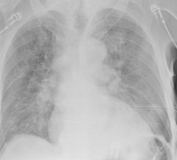
About 10 minutes later you are called to his room because he is complaining of dyspnea and his oxygen saturation has decreased to 86%. Breath sounds are somewhat decreased and the tube appears to be functioning properly. You immediately obtain another chest x-ray:

What just happened? This is a classic case of unilateral “flash” pulmonary edema after draining the chest cavity. This phenomenon was first described in 1853 in a patient who had just undergone thoracentesis. It is very uncommon, but seems to occur after rapid drainage of air or fluid from the chest cavity.

Here are some interesting factoids from case reports:

  • It occurs more often in young men
  • It is most common when draining large hemo- or pneumothoraces
  • Rapid drainage seems to increase the incidence
  • It is likely due to increased pulmonary capillary permeability from inflammatory mediators or changes in surfactant
  • Symptoms typically develop within an hour after drainage

What should you do? First, if you are draining a large collection of air or blood, do it slowly. Clamp the back end of the chest tube prior to insertion (you should always do this if you value your shoes) and use it to meter the amount of fluid or air released. I typically let out about 300cc of fluid, then wait a minute and repeat until all the blood has been drained. For air, vent it for 10 seconds, then wait a minute and repeat.

In patients at high risk for this condition, apply pulse oximetry and follow for about an hour. If they still look and feel great, nothing more need be done.

References:

  • Fulminant Unilateral Pulmonary Edema After Insertion of a Chest Tube. Dtsch Arztebl Int 105(50):878-881, 2008.
  • Reexpansion pulmonary edema after chest drainage for pneumothorax: A case report and literature overview. Respir Med Case Rep 14:10-12, 2015.
  • Re-expansion pulmonary edema following thoracentesis, Can Med Assn J 182(18):2000-2002, 2010.

Chest Tube Based On Pneumothorax Size

How big is too big? That has been the question for a long time as it applies to pneumothorax and chest tubes. For many, it is a math problem that takes into account the appearance on chest x-ray, the physiology of the patient, and their ability to tolerate the pneumothorax based on any pre-existing medical conditions.

I first wrote about this paper when it was just an abstract for last year’s AAST meeting. Apparently, it passed peer review muster. It has just been published in the Journal of Trauma. The numbers have changed a little bit, so I’ll update my analysis accordingly.

The group at Froedtert in Milwaukee has been trying to make the decision to place a chest tube a bit more objective. They introduced the concept of CT based size measurement using a 35mm threshold at the AAST meeting three years ago. Read my review here. My criticisms at the time centered around the need to get a CT scan for diagnosis and their subjective definition of a failure requiring chest tube insertion. The abstract never did make it to publication.

The authors are back now with a follow-on study. This time, they made a rule that any pneumothorax less than 35mm from the chest wall would be observed without tube placement. The performed a retrospective review of their experience and divided it into two time periods: 2015-2016, before the new rule, and 2018-2019, after the new rule. They excluded any chest tubes inserted before the scan was performed, those that included a sizable hemothorax, and patients placed on a ventilator or who died.

Here are the factoids:

  • There were 99 patients in the early period and 167 in the later period
  • Chest tube use significantly declined from 28% to 18% between the two periods. These numbers are 8% higher than were described in last year’s abstract.
  • Observation rates without a chest tube increased from 85% to 95% after implementation of the new guideline
  • There was no difference in length of stay, inpatient failure rate, complications, or death
  • The most common inpatient failure was due to development of a new hemothorax. However, there was an almost identical number of failures of “unclear” etiology. This is troublesome but part and parcel for such a retrospective study.
  • Two patients were readmitted within 30 days for a pulmonary complication (one empyema, one readmission at 3 days after discharge for dyspnea due to pneumothorax)
  • Patients in the later group were 2x more likely to be observed (by regression analysis)

The authors concluded that the 35mm rule decreased unnecessary chest tube insertion while maintaining patient safety.


Bottom line: I still have a few issues with this paper and the authors’ preceding series of abstracts. First, decision to insert a chest tube required a CT scan in a patient with a pneumothorax. This seems like extra radiation for patients who may not otherwise fit any of the usual blunt imaging criteria. And, like their 2018 and 2021 abstracts, there are no objective criteria for failure requiring tube insertion. So it is difficult to gauge compliance when insertion for failure is somewhat based on the whims of the individual surgeon.

What this abstract really shows is that compliance with the new rule increased, and there were no obvious complications from its use. The other numbers (chest tube insertions, observation failure) are just too subjective to learn much from. The most troubling issue is that the reason for 40% of failures was “unclear.” This is most likely due to the fact that the authors did not have objective guidelines for failure due to the retrospective nature of the study.

The numbers in this paper changed a little from last year’s abstract. The overall conclusions and meaning did not. It appears that 35mm is a reasonable threshold for pneumothorax size when contemplating inserting a chest tube. Unfortunately, this study relied entirely on CT scan. We don’t know if using a similar guideline for regular old chest x-ray is valid or not. 

What we still need is a good, prospective trial using an arbitrary guideline like 35mm pneumothorax as seen on chest x-ray or CT scan. And then, a clear definition of what defines a failure that requires tube insertion would be helpful. And at some point, we also need to know if a small tube or pigtail catheter is adequate for pure pneumothorax. Don’t get me started on that one!

Reference: The 35-mm rule to guide pneumothorax management: Increases appropriate observation and decreases unnecessary chest tubes. J Trauma 92(6):951-957, 2022.

Best Of EAST #11: Rib Fracture Fixation vs Epidural Analgesia

Rib fracture fixation has really taken off over the past five years for management of select rib fracture patterns. There are probably two mechanisms by which it improves pain control and speeds recovery.

The first is purely mechanical. In patients with flail chest, there is impairment of chest wall mechanics that decreases ventilatory efficiency and often leads to prolonged intubation and pulmonary complications. The other is the control of pain associated with multiple or displaced rib fractures.

The trauma group at the Brown University Alpert Medical School performed a TQIP database analysis that attempted to tease out the pain component in this equation. They compared outcomes from patients who underwent rib fixation or epidural analgesia within 72 hours of admission. They looked at a single year of TQIP data for adults with rib fractures, and excluded those who had TBI or died within 24 hours. Specific outcomes were pulmonary complications, lengths of stay, and mortality.

Here are the factoids:

  • There were just over 1,000 patients in each of the rib fixation and epidural analgesia groups
  • A much larger percentage of patients undergoing fixation had a diagnosis of flail chest (43% vs 13%) and a higher ISS (17 vs 14)
  • Early rib fixation was associated with an added 1.5 day length of stay, but this was not statistically significant
  • Early fixation was significantly associated with a higher risk of unplanned intubation
  • There were no differences in respiratory failure, VAP or mortality between the groups

The authors concluded that rib fracture fixation was associated with longer hospital length of stay but less risk of unplanned intubation. They suggest that patients should receive early referral to centers where both interventions are available so appropriate candidates can undergo fixation.

Bottom line: I’m struggling a bit here. When I read the title I thought I might learn something more about my therapeutic choices for patients with more complicated rib fractures. But this was not even a “how we did it” paper, but a “how hundreds of other centers did it” study. For a subject like this, a database study like this injects quite a bit of selection bias that just can’t be removed. 

For example, look at the huge (3x) difference in flail chest between the groups. Clearly, patients with a flail have a higher ISS and hospital length of stay, and are much more likely to selected for fixation. Thus, that diagnosis alone will skew the data more than the choice of procedure. I would suggest that simple descriptive and regression analyses is not adequate to answer your questions. Some type of propensity matching for ISS or at least AIS chest is probably required.

The only statistically significant result in the abstract was the decreased risk in unplanned intubation. Again, it’s difficult to say whether this is related to the larger percentage of patients who had flails who had their risk decreased by the procedure.

Here are my questions for the authors and presenter:

  1. Did you exclude all patients with TBI? Why not keep those with mild TBI (GCS 14-15), since they should behave similar to those without head injury?
  2. Why did you restrict your dataset to patients who underwent either procedure within the first 72 hours? This seems like an arbitrary time frame. Do you have a sense of the distribution of time interval until either procedure? As a thought experiment, let’s say that the mean (or median) time to either of the procedures was 5 days. You would be sampling the small, early tail of patients who had an intervention before day 3. In that case, your study might not be representative of of real life.
  3. Did you analyze the chest diagnoses and/or AIS  chest? Controlling or propensity score matching for this may have yielded additional information.
  4. You concluded that patients should be referred to centers where the best care can be provided. Isn’t this what we do already?

This is an interesting paper, and I’m hoping that you have more data to present than would fit in the abstract!

Reference: COMPARISON OF SURGICAL STABILIZATION OF RIB FRACTURES VS EPIDURAL ANALGESIA ON EARLY CLINICAL OUTCOMES. EAST 35th ASA, oral abstract #29.

Best Of EAST #5: Ultrasound vs Chest X-Ray After Chest Tube Removal

The chest is one of the most commonly injured body regions. Patients are frequently found to have either air or blood in the chest, and many require a chest tube (tube thoracostomy) for these conditions. There is an art to chest tube removal, and even in 2021, the best practice has not been fully worked out.

Some believe that pulling the tube during a breath hold is best. Others do this during full expiration. Most centers confirm an uneventful tube removal with a plain chest x-ray. But the time interval after removal varies considerably.

The trauma group at the University of Tennessee – Chattanooga examined the use of chest ultrasound as the confirmatory test for residual pneumothorax after removing a chest tube. They developed an institutional practice guideline requiring a trans-thoracic ultrasound performed by a first-year resident two hours after tube removal. The interns all completed a 30-minute standard ultrasound course for training prior to beginning the study.

Two hours after tube removal, an intern performed the ultrasound (US) and interpreted it. A chest x-ray (CXR) was then ordered and the results compared.

Here are the factoids:

  • A retrospective review of 46 patients was performed, but the inclusion criteria were not listed in the abstract
  • Eleven of the 46 (24%) had a residual pneumothorax on CXR, and the US detected it in 12 (26%)
  • Three patients had PTX on CXR, but not US
  • Four patients had PTX on US, but not CXR
  • None of the PTX were clinically significant, and none required tube reinsertion
  • Cost of care savings was projected to be $4,000 if chest x-ray was not needed

The authors concluded that bedside ultrasound was an acceptable alternative to chest x-ray, with decreased radiation exposure and cost.

Bottom line: This is an intriguing abstract. It shows us that there might be an alternative to the standard chest x-ray confirmation after chest tube removal. It’s a very small study, so non-inferiority can’t truly be established yet. The studies are complementary since each study misses a few pneumothoraces that the other picks up.

At this point, I wouldn’t recommend switching entirely to ultrasound until we have a larger series. But I bet we will be able to in the future. Ultimately, this could reduce radiation exposure (tiny anyway for a chest x-ray) and save a small amount of money. But it will reduce x-ray department resource usage, which may be very helpful for the hospital.

Here are my questions for the authors and presenter:

  • How did you select your patients? What were the selection criteria? How long did it take to accrue 46 patients? It’s important that all patients with a chest tube had the criteria applied, otherwise there is an opportunity for bias. We want to make sure that you didn’t inadvertently enroll only the patients for whom ultrasound works well.
  • How much of a burden was placed on the interns who did the exam? Was the ultrasound unit nearby? Or did they have to spend 30 valuable minutes rolling it to the floor and doing the study? Radiology department resource use needs to be balanced with intern resource utilization.
  • Why did you have such a high rate of residual pneumothorax after the tubes were pulled (about 25%). This seems a bit higher than what the literature reports.
  • What does your protocol require when a residual pneumothorax is found? Do you have to perform another study after an additional time interval to prove that it is not getting larger? Serial ultrasound exams? Another chest x-ray? Please show us your entire guideline.

I really enjoyed this paper. I’m looking forward to hearing the nitty gritty details during the presentation.

Reference: ULTRASOUND SAFELY REPLACES CHEST RADIOGRAPH AFTER TUBE THORACOSTOMY REMOVAL IN TRAUMA PATIENTS. EAST 25th ASA, oral abstract #9.

Retained Hemothorax: The Practice Guideline

Over the last few days, I’ve reviewed some data on managing hemothorax, as well as the use of lytics. Then I looked at a paper describing one institution’s experience dealing with retained hemothorax, including the use of VATS. But there really isn’t much out there on how to roll all this together.

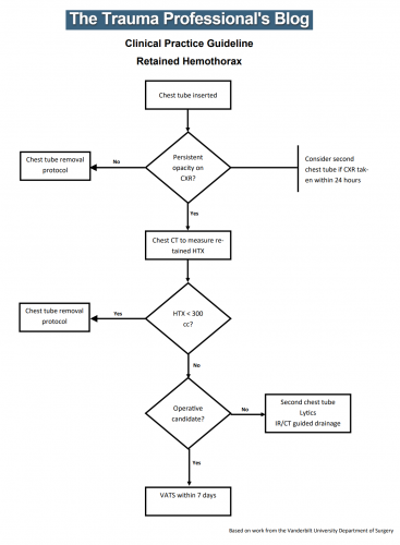
Until now. The trauma group at Vanderbilt published a paper describing their experience with a home-grown practice guideline for managing retained hemothorax.  Here’s what it looks like:

I know it’s small, so just click it to download a pdf copy. I’ve simplified the flow a little as well.

All stable patients with hemothorax admitted to the trauma service were included over a 2.5 year period. The practice guideline was implemented midway through this study period. Before implementation, patients were treated at the discretion of the surgeon. Afterwards, the practice guideline was followed.

Here are the factoids:

  • There were an equal number of patients pre- and post-guideline implementation (326 vs 316)
  • An equal proportion of each group required an initial intervention, generally a chest tube (69% vs 65%)
  • The number of patients requiring an additional intervention (chest tube, VATS, lytics, etc) decreased significantly from 15% to 9%
  • Empyema rate was unchanged at 2.5%
  • Use of VATS decreased significantly from 8% to 3%
  • Use of catheter guided drainage increased significantly from 0.6% to 3%
  • Hospital length of stay was the same, ranging from 4 to 11 days (much shorter than the lytics studies!)

Bottom line: This is how design of practice guidelines is supposed to work. Identify a problem, typically a clinical issue with a large amount of provider care variability. Look at the literature. In general, find it of little help. Design a practical guideline that covers the major issues. Implement, monitor, and analyze. Tweak as necessary based on lessons learned. If you wait for the definitive study to guide you, you’ll be waiting for a long time.

This study did not significantly change outcomes like hospital stay or complications. But it did decrease the number of more invasive procedures and decreased variability of care, with the attendant benefits from both of these. It also dictates more selective (and intelligent) use of additional tubes, catheters, and lytics. 

I like this so much that I’ve incorporated parts of it into the chest tube guideline at my center!

Download the practice guideline here.

Related posts:

Reference: Use of an evidence-based algorithm for patients with traumatic hemothorax reduces need for additional interventions. J Trauma 82(4):728-732, 2017.