Category Archives: Imaging

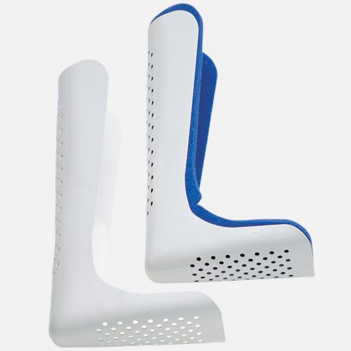
Part 2: Metal Splints – Can You CT Scan Through Them?

In my last post, I debunked the myth that using a pre-formed aluminum splint significantly degrades the quality of standard x-rays. But what about a study that provides much more detail, such as CT scan?

CT scan techs have told me that there would be too much artifact using any kind of metal splint. And typically, when imaging an extremity with CT, we are looking at vascular runoff. The vessels are small, and high image quality is extremely important. If the images are bad, then we risk having to give the patient another dose of both radiation and contrast.

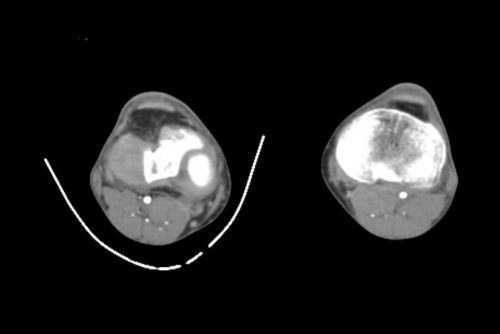
As you know, my mantra is question everything! So i scouted around and found some images to share using one of these splints. Look closely for the intimal flap in the image below:

Can’t see it? That’s because it isn’t there! But you certainly could if it were!

Bottom line: A perforated aluminum splint causes absolutely no artifact or image degradation. Do not cause additional injury by removing it prior to imaging, either CT or conventional x-ray. Although your friendly techs, radiologists, and orthopedic surgeons may moan, it won’t hurt their ability to make decisions on the images.

Part 1: Metal Splints – Can You X-ray Through Them?

Splinting is an important part of the trauma resuscitation process. No patient should leave your trauma resuscitation room without splinting of all major fractures. It reduces pain, bleeding, and soft tissue injury, and can keep a closed fracture from becoming an open one.

But what about imaging? Can’t the splint degrade x-rays and hamper interpretation of the fracture images? Especially those pre-formed aluminum ones with the holes in them? It’s metal, after all.

Some of my orthopedic colleagues insist that the splint be removed in the x-ray department before obtaining images. And who ends up doing it? The poor radiographic tech, who has no training in fracture immobilization and can’t provide additional pain control on their own.

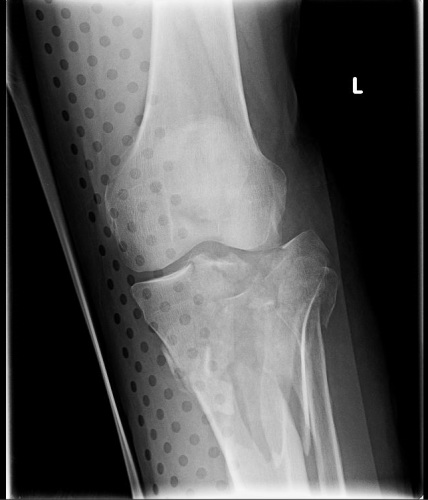
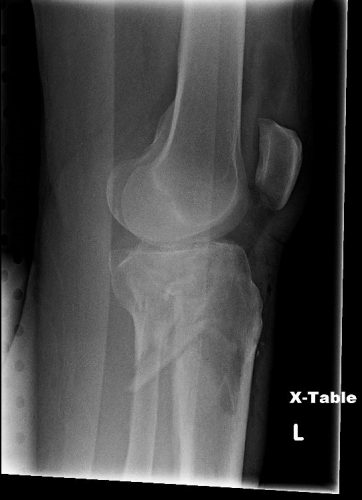
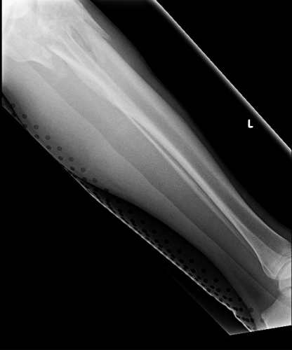
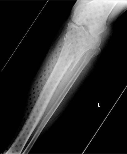
But does it really make a difference? Judge for yourself. Here are some knee images with one of these splints on:

Amazingly, this thin aluminum shows up only faintly. There is minimal impact on interpretation of the tibial plateau. And on the lateral view, the splint is well posterior to bones.

On the tib-fib above, the holes are a little distracting on the AP view, but still allow for good images to be obtained.

Bottom line: In general, splints should not be removed during the imaging process for acute trauma. For most fractures, the images obtained are more than adequate to define the injury and formulate a treatment plan. If the fracture pattern is complex, it may be helpful to temporarily remove it, but this should only be done by a physician who can ensure the fracture site is handled properly. In some cases, CT scan may be more helpful and does not require splint removal. And in all cases, the splint should also be replaced immediately at the end of the study.

In my next post, I’ll look at the use of CT scans when this type of splint is in use.


How Much Radiation Exposure In Imaging Studies?

Everyone knows that CT scans deliver more radiation than conventional x-ray. But how much does each test really deliver? And how significant is that?

Let me try to put it all into perspective. First, how much radiation are we exposed to just living outside the hospital? Background radiation is everywhere. It consists of radioactive gases (argon) in the air we breathe, radiation from the rocks and other things around us, and cosmic rays blasting through us from space.

In the United States, the average background radiation each of us is exposed to is about 3.1 milliSieverts (mSv). I’ve compiled a table to show the approximate dose delivered by some of the common radiographic studies ordered by trauma professionals. And to keep it real, I’ve calculated how much extra background radiation we would have to absorb, in units of time, to have an equivalent exposure.

Read and enjoy! Remember, doses may vary by scanner, settings, and dose reduction measures used.

Test Dose (mSv) Equivalent background
radiation
Chest x-ray 0.1 10 days
Pelvis x-ray 0.1 10 days
CT head 2 8 months
CT cervical spine 3 1 year
Plain c-spine 0.2 3 weeks
CT chest 7 2 years
CT abdomen/pelvis 10 3 years
CT T&L spine 7 2 years
Plain T&L spine 3 1 year
Millimeter wave
scanner (that hands
in the air TSA thing at
the airport)
0.0001 15 minutes
Scatter from a chest
x-ray in trauma bay
when standing one 
meter from the
patient
0.0002 45 minutes
Scatter from a chest
x-ray in trauma bay
when standing three 
meters from the
patient
0.000022 6 minutes

ACS Trauma Abstract #6: Scanning Unstable Trauma Patients???

If you’ve read my stuff for very long, you know I frown on sending unstable patients anywhere but to the OR. Instability tends to get worse, and that always happens at inopportune locations like hallways, elevators, and CT scanners. Imagine my surprise when I noticed an abstract being presented at the Clinical Congress of the American College of Surgeons this week suggesting that it was okay to scan hemodynamically unstable patients before “definitive therapy.”

Here’s the title:

“Computed tomography in hemodynamically abnormal thoracoabdominal trauma safely enhances surgical triage”

The devil is in the details and the language. This group from USC included all patients who were hemodynamically abnormal on arrival to the trauma bay but who normalized to SBP > 90 during the resuscitation were included.  A total of 253 of these patients were reviewed over a 9 year period, and the usual variables were analyzed (mortality, complications, hospital, ICU, and vent days, etc).

Here are the factoids:

  • Of the 253 patients studied, 45 went to straight to OR and 208 were taken to CT
  • Injury severity was identical for the two groups
  • Lengths of stay and mortality were not different, but only p values were given
  • Patients taken to CT cleared their lactic acidosis faster (12 vs 5 hours), and used a bit less plasma and significantly less blood transfusions
  • The OR group underwent more procedures (31% vs 13%), although what these were and when they were performed is not listed

Bottom line: The title of this abstract is misleading, and may fool those who don’t read the rest of the abstract. It should read:

“Computed tomography in previously hemodynamically abnormal thoracoabdominal trauma safely enhances surgical triage”

Someone who just skims through this issue of the journal may get the idea that it’s okay to scan an unstable patient. The authors are not saying this at all. If you read the conclusion carefully, you can see that the patients had to be resuscitated to a SBP > 90 before they considered taking to scan. And they did that for the majority of these patients.

The real question is, why do the scanned patients clear their lactic acidosis faster, need less blood, and undergo fewer procedures? It appears that there is some bias or selection process in play. Otherwise, why not use the magic CT scanner to make them all better?

Reference: Computed tomography in hemodynamically abnormal thoracoabdominal trauma safely enhances surgical triage. JACS 225(4S2):e175-176, 2017.