Category Archives: General

What Does A Retained Surgical Sponge Look Like?

Surgeons and surgical residents rarely see these. And because it’s so uncommon, they frequently don’t recognize the telltale findings on radiographic studies. The TSA runs into the same problem in screening passengers for weapons and other hazards at airports. But it’s the bane of any surgeon’s existence. And it’s a major reason why OR personnel take such great pains to account for everything in the room. It is a catastrophe, and always a preventable one, when some piece of equipment goes missing and ends up left inside a patient.

A number of methods have been developed to try to eliminate this problem. They include careful counts, having someone record anytime anything is placed inside, x-rays, and most recently, RFID tags.

After counting, x-ray is the most common way to try to find missing objects. One would think that these foreign bodies would be easy to see. Metallic instruments are rather easy to spot. But many trauma professionals, even those who work in the OR, have never seen what a positive image of a sponge actually looks like. So here they are. You should never miss one on an xray now.

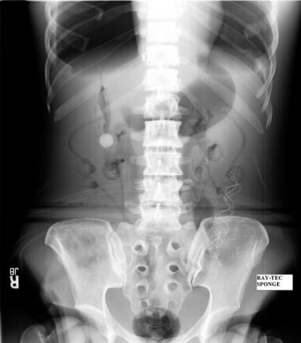
Surgeons typically use two types of sponges in the OR: Ray-Tec sponges and standard lap pads. Ray-Tecs look like a 4×8 piece of gauze with a mysterious blue string woven throughout it. The string is the only part that shows up on x-ray, and it is very thin and somewhat hard to see. Here are some Ray-Tec sponges outside the body:

And here’s one that was left inside. Note the little squiggle in the left lower quadrant and how easy it is to overlook.
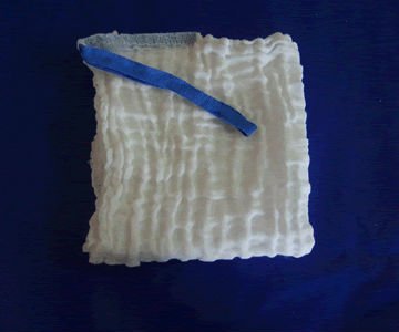
On the other hand, a laparotomy pad is a 4×4 folded cloth pad that unfolds into a larger pad. It has a blue tag sewn in the corner, extending along one edge of the pad. Here’s what they look like outside the body:
And here’s one inside a patient. Note the irregular object in the right upper quadrant.
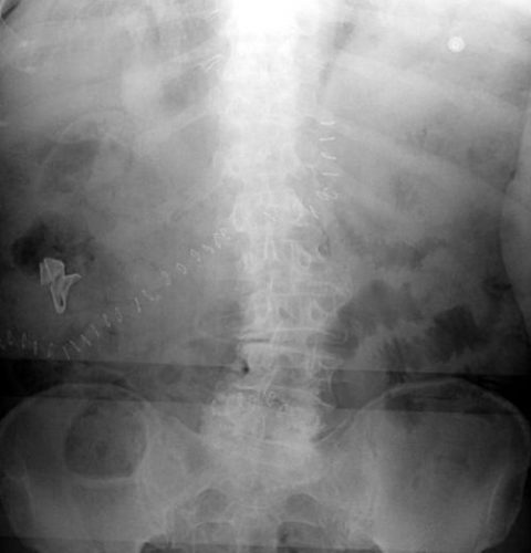
Bottom line: It’s important for anyone who works in the OR on any body part to be familiar with the appearance of these tags on x-rays. Since it’s generally impossible to get accurate counts before or after a trauma procedure, always image the involved body cavity looking for these telltale signs before closing the patient.

Note: These images taken from the internet. Patients not treated at Regions Hospital.

Print Friendly, PDF & Email

Gunshots And CT Scan Of The Abdomen

Abdominal gunshots and CT scanning are usually thought to be mutually exclusive. The usual algorithm generally means a prompt trip to the operating room. But as with many things in the management of trauma, there are always exceptions. The key is to understand when exactly one of those exceptions is warranted.

Exception 1: Did it really enter the abdomen? Gunshots have enough energy that they usually do get inside. However, freaky combinations of trajectory and body habitus do occur. There are three tests that must be passed in order to entertain the possibility that the bullet may not have made it inside your patient: physiology, anatomy, and physical exam. For physiology, the patient must be completely hemodynamically stable. Anatomically, the trajectory must make sense. If the known wounds and angles allow a tangential course make sense, then fine. But if there is a hole in the epigastrium and another next to the spine, you have to assume the bullet went straight through. Finally, the physical exam must be normal. No peritonitis. No generalized guarding. Focal tenderness only in the immediate area of any wounds. If all three of these criteria are passed, then a CT can be obtained to demonstrate the trajectory.

Exception 2: Did it enter an unimportant area of the abdomen? Well, there’s really only one of these, and that’s the area involving the right lobe of the liver and extending posteriorly and lateral to it. If the bullet hole(s) involve only this area, and the three tests above are passed, CT may confirm an injury that can be observed. However, there should only be a minimal amount of free fluid, and no soft tissue changes of any kind adjacent to bowel.

Exception 3: A prompt trauma lap was performed, but you think you need more information afterwards. This is rare. The usual belief is that the eyes of the surgeon provide the gold standard evaluation during a trauma lap. For most low velocity injuries with an easily understood trajectory, this is probably true. However, high velocity injuries, those involving multiple projectiles, or complicated trajectories (side to side) can be challenging for even the most experienced surgeon. Some areas (think retroperitoneum or deep in the pelvis) are tough to visualize completely, especially when there’s blood everywhere. These are also the cases most likely to require damage control surgery, so once the patient has been temporarily closed, warmed and resuscitated, a quick trip to CT may be helful in revealing unexpected shrapnel, unsuspected injuries, or other issues that may change your management. Even a completely unsurprising scan can provide a higher sense of security.

Bottom line: CT of the abdomen and gunshots to that area may actually coexist in some special cases. Make sure the physiology, anatomy and physical exam criteria are passed first. I also make a point of announcing to all trainees that taking these patients to CT is not the norm, and carefully explain the rationale. Finally, apply the concept of the null hypothesis to this situation. Your null hypothesis should state that your patient does not need a CT after gunshot to the abdomen, and you have to work to prove otherwise!

Print Friendly, PDF & Email

Practice Guidelines And Tincture Of Time

Most trauma centers have at least a few practice guidelines to help the standardize the way they manage common injuries. Solid organ injury. Elder trauma. Chest tube management. But they are all designed for use in patients who present shortly after their injury.

What about someone who presents a day or two, or more, after their injury?  That changes the picture entirely. Most guidelines have a time component built in. A TBI protocol requires a repeat head CT after a certain period of time. Solid organ injury patients may have restricted activity or frequent vital signs for a while. 

But all too often, trauma professionals treat the patient with delayed presentation exactly the same as fresh trauma. For example, a patient falls and bumps their head. They have a persistent headache, and after two days decide to visit their local ED. The CT scan shows a small amount of subarachnoid blood in the area of the impact. Your practice guidelines says to admit for observation, frequent neruo checks, and repeat head CT in 12 hours.

Or a young male playing sports took a hit to his left flank. After 3 days, he’s just tired of the pain and comes to the ED for some pain medication. CT scan shows a grade III spleen injury with a small amount of hemoperitoneum. Your protocol says to admit, make NPO, liimit activity, and observe for 2 days.

What would I do in these cases? Think about it! If the patients had presented right after the event, they would have gone through your guideline and would have been discharged already. So I would review the images, talk to the patients about their injuries, then send them home from the ED with followup. They’ve already passed!

Bottom line: Remember, practice guidelines are not etched in stone. Variances are possible, but need to be well thought out in advance. And hopefully documented in the chart to expedite the inevitable trauma performance improvement inquiry. If the requisite amount of time has gone by, and the history and exam are reasonable, the patient has already passed your protocol. Send them home.

Related posts:

Print Friendly, PDF & Email

The Fifth Law Of Trauma – Pediatric

I knew there was a fifth law! Any time I give a pediatric talk, I mention it. This one applies to anyone who takes care of children, and is particularly important to EMS / prehospital providers and emergency physicians.

On occasion, medics are called to a home to treat a child in extremis, or occasionally in arrest. Similarly, extremely sick children are often brought to the ED by parents or other caregivers.

Here’s the fifth law:

A previously healthy child who is in arrest, or nearly so, is a victim of child abuse until proven otherwise.

Bottom line: It’s so easy to go down the sepsis path with sick kids, especially those who can’t talk yet. But healthy children tend to stay healthy, and don’t easily get sick to the point of physiologic collapse. If you encounter one as a prehospital provider, glance around at the environment, and evaluate the caregivers. In the ED, ask pointed questions about the circumstances and do a full body examination. What you hear and what you see may drastically alter how you evaluate the patient and may save their life.

Other Laws of Trauma:

Print Friendly, PDF & Email

Prehospital To Trauma Team Handoff: A Solution

I wrote about handoffs between EMS and the trauma team yesterday. It’s a problem at many hospitals. So what to do?

Let’s learn from our experience in the OR. Best practice in the operating room mandates a specific time out process that involves everyone in the OR. Each participant in the operation has to stop, identify the patient, state what the proposed procedure and location is, verify that the site is marked properly, and that they have carried out their own specific responsibilities (e.g. infused the antibiotic).

Some trauma centers have initiated a similar process for their trauma team as well. Here’s how it works:

  • The patient is rolled into the resuscitation room by EMS personnel, but remains on the stretcher.
  • Any urgent cares continue, such as ventilation.
  • The trauma team leader is identified and the EMS lead gives a brief report while everyone in the room listens. The report consists of only mechanism, all identified injuries, vital signs (including pupils and GCS), any treatments provided. This should take no more than 30 seconds.
  • An opportunity for questions to be asked and answered is presented
  • The patient is moved onto the hospital bed and evaluation and treatment proceed as usual.
  • EMS personnel provide any additional information to the scribe, and may be available to answer any additional questions for a brief period of time.

Bottom line: This is an excellent way to improve the relationship between prehospital and trauma team while improving patient care. It should help increase the amount of clinically relevant information exchanged between care providers. Obviously, there will be certain cases where such a clean process is not possible (e.g. CPR in progress). I recommend that all trauma programs consider implementing this “Trauma Activation Time Out For EMS” concept.

Tomorrow, I’ll share a best practice to make this process even better!

Related posts:

image
Print Friendly, PDF & Email