Category Archives: General

The Right Way to Treat Tension Pneumothorax

Tension pneumothorax is an uncommon but potentially lethal manifestation of chest injury. An injury to the lung occurs that creates a one-way valve effect, allowing a small amount of air to escape with every breath. Eventually the volume becomes so large as to cause the lung and mediastinum to push toward the other side, with profound hypotension and cardiovascular collapse.

The classic clinical findings are:

  • Hypotension
  • Decreased or absent breath sounds on the affected side
  • Hyperresonance to percussion
  • Shift of the trachea away from the affected side
  • Distended neck veins

You should never diagnose a tension pneumothorax with a chest xray or CT scan, because the diagnosis is a clinical one and the patient may die while these procedures are carried out. Having said that, here’s one:

Tension Pneumothorax

The arrow points to the completely collapsed lung. Note the trachea bowing to the right. 

As soon as the diagnosis is made, the right thing to do is to “needle the chest.” A large bore angiocath should be placed in the second intercostal space, mid-clavicular line, sliding right over the top of the third rib. The needle should then be removed, leaving the catheter.

The traditional large bore needle is 14 gauge, but they tend to be short and flimsy. They may not penetrate the pleura in an obese patient, and will probably kink off rapidly. Order the largest, longest angiocath possible and stock them in your trauma resuscitation rooms.

image

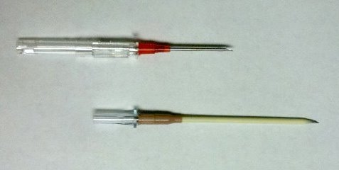
The top catheter in this photo is a 14 gauge 1.25 inch model. The bottom (preferred at Regions) is a 10 gauge 3 inch unit. Big difference! And if the patient is extremely obese, make a 1 cm cut in the skin and sink the hub deep to the skin for extra distance.

The final tip to treating a tension pneumothorax is that a chest tube must be placed immediately after inserting the needle. If the patient is on a ventilator, the positive pressure will slowly expand the lung. But if they are breathing spontaneously, the needle will change the tension pneumothorax into a simple open pneumothorax. Patients with other cardiovascular problems will not tolerate this for long and may need to be intubated if you dawdle.

Print Friendly, PDF & Email

Management of CSF Otorrhea/Rhinorrhea

The management of CSF leaks after trauma remains somewhat controversial. The literature is sparse, and generally consists of observational studies. However, some general guidelines are supported by large numbers of retrospectively reviewed patients.

  • Ensure that the patient actually has a CSF leak. In most patients, this is obvious because they have clear fluid leaking from ear or nose that was not present preinjury. Here are the options when the diagnosis is less obvious (i.e. serosanguinous drainage):
    • High resolution images of the temporal bones and skull base. If an obvious breach is noted, especially if fluid is seen in the adjacent sinuses, then a CSF leak is extremely likely.
    • Glucose testing. CSF glucose is low compared to serum glucose. 
    • Beta 2 transferrin assay. This marker is very specific to CSF. However, the test is expensive and results may take several days to a few weeks to receive. Most leaks will have closed before the results are available, making this a poor test.
  • Place the patient at bed rest with the head elevated. The basic concept is to decrease intracranial pressure, which in turn should decrease the rate of leakage. This same technique is used for management of mild ICP increases after head injury.
  • Consider prophylactic antibiotics carefully. The clinician must balance the likelihood of meningitis with the possibility of selecting resistant bacteria. If the likelihood of contamination is low and the patient is immunocompetent, antibiotics may not be needed.
  • Ear drops are probably not necessary. They may confuse the picture when gauging resolution of the CSF leak.
  • Wait. Most tramatic leaks will close spontaneously within 7-10 days. If it does not, a neurosurgeon or ENT surgeon should be consulted to consider surgical closure.

References:

  1. Brodie HA, Thompson TC. Management of complications from 820 temporal bone fractures. Am J Otol, 1997;18:188-197.
  2. Brodie HA. Prophylactic antibiotics for posttraumatic cerebrospinal fluid fistulas. Arch Otolaryngol Head, Neck Surg. 123:749-752.
Print Friendly, PDF & Email

Trauma Team Qualifications

What kind of credentialing should the members of your trauma team have?

The most important concept in answering this question is based purely on patient care. What shows that your personnel can provide the type of care that their position on the team warrants?

For all physicians, ATLS is a must. Verification agencies, including the American College of Surgeons, require that most providers to have passed the course once. I recommend a current ATLS provider certificate for all physicians, including residents. The course is updated every four years, which means that every time a provider certificate expires, new course content has been added. A current certificate keeps their knowledge up to date.

The airway physician should be either credentialed or have generous experience in managing the airway. This ensures that the intubation process is routine and safe when necessary.

Most, if not all physicians should be also current in ACLS, since major trauma patients may arrest prior to or after arrival at the hospital.

Nurses involved in trauma resuscitations should have additional credentialing such as TNCC. They should also be current in ACLS, and many hospital have added critical care experience and ongoing training requirements related to trauma.

For pediatric trauma programs, a basic pediatric resuscitation course such as PALS or APLS is mandatory. All physicians and nurses taking care of critically injured children should be current so they can provide the best care possible to them.

A piece of paper does not necessarily prove competence. The final part of the qualification process involves your trauma Performance Improvement program. All providers are scrutinized by the PI process, and if any stand out as having too many adverse events to their name, they should not be allowed to participate on the team.

The bottom line: Your trauma team members need to be great at what they do, with appropriate paperwork to prove it and PI monitoring to back it up. They need to be able to easily pass the “family member” test: would I want this person to take care of my (daughter) (husband) (grandmother) etc!

Print Friendly, PDF & Email

Teaching the Trauma Team

Teaching hospitals have extra responsibilities when constructing their trauma activation team. They are typically charged with educating trainees from a variety of disciplines, including residents, medical students, and students from other disciplines (EMT, PA, NP). The activation process must not only provide rapid and high quality trauma care, it must also teach these students how to provide that care.

Residents can be integrated into the typical physician roles on the team: airway and primary examiner. To integrate more trainees, these roles can be split further. For example, the examiner’s role can be split into a primary examiner and a secondary examiner with separate, lesser responsibilities. PAs and NPs can be integrated into these roles as well.

One of the most important “additions” to the team that allows education of senior level residents is the Team Leader. This role allows the trainee to learn how to direct the overall resuscitation and allows them to practice making management decisions on the fly. Typically, the Team Leader does not actually touch the patient, allowing the other examining physicians to do this and learn their specific roles. Each role can be assigned to an appropriate level resident, so that they move to higher levels as they progress through their training program.

Here is a template for a trauma team that allows four trainees (yellow balloons) to participate. One faculty members supervises all of them.

At our Trauma Center, we have these four trainees plus another Emergency Medicine resident who performs the FAST exam, if indicated. Two faculty members participate, one trauma surgeon and one Emergency Medicine faculty. Our total team size is 12, so it must be well-coordinated in order to avoid chaos.

Medical and paramedic students are usually confined to the periphery to take notes (H&P) or just observe.

Please leave your comments describing the composition of your team and what makes it run well.

Tomorrow: qualifications of your trauma team personnel

Print Friendly, PDF & Email