Category Archives: Complications

A Sample Final Damage Control X-ray

Yesterday, I wrote about ways to reduce and hopefully eliminate retained foreign bodies (instruments, sponges) during damage control surgery. Today, I’ll provide a sample x-ray and some tips on how to use this tool most effectively.

Here is an abdominal x-ray obtained just prior to closure of a patient who underwent damage control laparotomy. The OR record and surgeon from the initial operation documented that four sponges had been left in place for hemostasis.

dc-closure

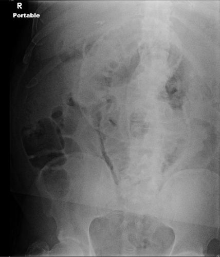
Nothing retained, right?

Wrong! This image is not complete. This patient is larger than the x-ray plate used. The area under the diaphragms, the pelvis, and the entire left side of the peritoneal cavity have not been visualized.

Tips for imaging for damage control closure:

  • Always make sure the patient is on an x-ray OR table. It is so annoying (and potentially a sterility problem) to have to slide the plate under the patient!
  • Help the radiology tech to locate the desired imaging field using folds in the towels covering the body region. For example place the confluence of folds in the center of the towel in the exact place you want the center of the x-ray to be.
  • Remove all radiopaque objects from the x-ray field to reduce confusion when interpreting the image
  • Make sure the entire body cavity has been imaged! This may mean bracketing the area with several shots.
  • Read the image yourself! But if in doubt, or in patients with drains or other odd objects, call the radiologist to help you out.

Related posts:

The Final X-Ray In Damage Control Surgery

Damage control surgery for trauma is over 20 years old, yet we continue to find ways to refine it and make it better. Many lives have been saved over the years, but we’ve also discovered new questions. How soon should the patient go back for definitive closure? What is the optimal closure technique? What if it still won’t close?

One other troublesome issue surfaced as well. We discovered that it is entirely possible to leave things behind. Retained foreign bodies are the bane of any surgeon, and many, many systems are in place to avoid them. However, many of these processes are not possible in emergent trauma surgery. Preop instrument counts cannot be done. Handfuls of uncounted sponges may be packed into the wound.

I was only able to find one paper describing how often things are left behind in damage control surgery (see reference below), and it was uncommon in this single center study (3 cases out of about 2500 patients). However, it can be catastrophic, causing sepsis, physical damage to adjacent organs, and the risk of performing an additional operation in a sick trauma patient.

So what can we do to reduce the risk, hopefully to zero? Here are my  recommendations:

  • For busy centers that do frequent laparotomy or thoracotomy for trauma and have packs open and ready, pre-count all instruments and document it
  • Pre-count a set number of laparotomy pads into the packs
  • Use only items that are radiopaque or have a marker embedded in them. This includes surgical towels, too!
  • Implement a damage control closure x-ray policy. When the patient returns to OR and the surgeons are ready to begin the final closure, obtain an x-ray of the entire area that was operated upon. This must be performed and read before the closure is complete so that any identified retained objects can be removed.

Tomorrow, a sample damage control closure x-ray.

Related post:

Reference: Retained foreign bodies after emergent trauma surgery: incidence after 2526 cavitary explorations. Am Surg 73(10):1031-1034, 2007.

Predicting VTE Risk In Children

There’s a lot of debate about if and at what age injured children develop significant risk for venous thromboembolism (VTE). In the adult world, it’s a little more clear cut, and nearly every patient gets some type of prophylactic device or drug. Kids, we’re not so certain about at all.

The Children’s Hospital of Wisconsin tried to tease out these factors to develop and implement a practice guideline for pediatric VTE prophylaxis. They prospectively reviewed over 4000 pediatric patients admitted over a 6 year period.

It looks like the guideline was developed using some or all of this data, then tested using regression models to determine which factors were significant. The guideline was then tweaked and a final model implemented.

Here are the factoids:

  • 588 of the patients (14%) were admitted to the ICU, and 199 of these were identified as high risk by the guidelines
  • Median age was 10 (this is always important in these studies)
  • VTE occurred in 4% of the ICU patients, and 10% of the high risk ones
  • Significant risk factors included presence of central venous catheter, use of inotropes, immobilization, and GCS < 9

Bottom line: This abstract confuses me. How were the guidelines developed? What were they, exactly? And the results seem to pertain to the ICU patients only. What about the non-ICU kids? The abstract just can’t convey enough information to do the study justice. Hopefully, the oral presentation will explain all.

I prefer a very nice analysis done at the Oregon Health Science University in Portland. I wrote about this study earlier this year. The authors developed a very useful calculator that includes most of the risk factors in this model, and a few more. Input the specific risks, and out comes a nice score. The only issue is, what is the score threshold to begin prophylaxis and monitoring? Much more practical (and understandable) than this abstract. Check it out at the link below.

Related post:

References:

  1. Evaluation of guidelines for injured children at high risk for VTE: a prospective observational study. AAST 2016, Paper 68.
  2. A Clinical Tool for the Prediction of Venous Thromboembolism in Pediatric Trauma Patients. JAMA Surg 151(1):50-57, 2016.

How To Predict Venous Thromboembolism In Pediatric Trauma

As with adults a decade ago, the incidence of venous thromboembolism (VTE) in children is now on the rise. Whereas adult VTE occurs in more than 20% of adult trauma patients without appropriate prophylaxis, it is only about 1% in kids, but increasing. There was a big push in the early 2000′s to develop screening criteria and appropriate methods to prevent VTE. But since the incidence in children was so low, there was no impetus to do the same for children.

The group at OHSU in Portland worked with a number of other US trauma centers, and created some logistic regression equations based on a large dataset from the NTDB. The authors developed and tested 5 different models, each more complex than the last. They ultimately selected a model that provided the best fit with the fewest number of variables.

The tool consists of a list of risk factors, each with an assigned point value. The total point value is then identified on a chart of the regression equation, which shows the risk of VTE in percent.

Here are the factors:

Note that the highest risk factors are age >= 13, ICU admission, and major surgery.

And here is the regression chart:

Bottom line: This is a nice tool, and it’s time for some clinical validation. So now all we have to do is figure out how much risk is too much, and determine which prophylactic tools to use at what level. The key to making this clinically usable is to have a readily available “VTE Risk Calculator” available at your fingertips to do the grunt work. Hmm, maybe I’ll chat with the authors and help develop one!

Reference: A Clinical Tool for the Prediction of Venous Thromboembolism in Pediatric Trauma Patients. JAMA Surg 151(1):50-57, 2016.

Post-Embolization Syndrome?

Important! If your search for a trauma topic brought you here…

The Trauma Professional’s Blog has moved to this new site, but old Google or Bing searches may link you to this page.

Please retype your search in the box in the upper left corner of any page (or touch the magnifying glass above on a mobile device) to find what you are looking for.

Go to theTrauma Professional's Blog page

And hopefully, all the search engines will finish indexing the new site soon! Explore the site and enjoy! Michael

———————————————————————————-

I’ve seen a number of trauma patients who have developed pain and elevated WBC after embolization of solid organs for trauma. For kidneys and main splenic artery embolization, it’s fairly common in my experience. Turns out, this phenomenon was described in 2007-2008 in patients undergoing embolization of hepatic tumors and uterine fibroids. It was termed post-embolization syndrome, and consists of pain, fever, nausea and ileus.

An article was just published in the Journal of Trauma describing this syndrome in children after splenic embolization for blunt trauma. The authors looked at their own trauma registry over a 12 year period. Yes, it took that long to find 448 children with blunt splenic injury. Of those, only 11 underwent arterial embolization (sigh of relief).

The average age was about 13 and ISS was 16 in both groups. Kids who underwent embolization were more likely to spend some time in the ICU, had a longer hospital stay (8 vs 5 days(!)), and took longer to resume their diet (5 vs 2 days). These differences occurred despite the fact that most of the embolized children had isolated splenic injuries. Additionally, the embolized children were more likely to receive blood (3 units vs none) and plasma.

My first question about this paper is, why? Broken spleens in children do not act like broken spleens in adults. The vast majority of the cases of contrast extravasation in children stops on its own without intervention. So why did we even have to find out that post-embolization syndrome occurs in children? They shouldn’t be going through this procedure anyway! Fortunately, a deeper read of the paper provides the answer. The indication for angio was splenic pseudoaneurysm in 2, and ongoing hemorrhage in the other 9. In the case of these latter 9, it did keep the children from having their spleens operated on.

Bottom line: In general, don’t send kids for splenic angiography (99.3% of kids in this study did not have it). Ongoing hemorrhage (prior to hypotension, which is an absolute indication for OR) is probably the only indication I can think of. Pseudoaneurysm and extravasation of contrast are not indications like they are in adults. But if you do have to send them, just be aware that they may develop pain, fever and ileus that will keep them in the hospital and/or ICU for a few extra days.

Reference: Transarterial embolization in children with blunt splenic injury results in postembolization syndrome: A matched case-control study. J Trauma 73(6):1558-1563, 2012.