Category Archives: Complications

Incidental Finding: Gas In The Spleen After Embolization

Most solid organ injury practice guidelines include angioembolization in part of the pathway. But very few require re-imaging at any point to see how the liver or spleen are coming along.

But every once in a while another condition arises, or symptoms worsen unexpectedly, causing us to get another CT scan that includes the abdomen and pelvis. And sometimes we see things that we wouldn’t normally see, like air bubbles in the organ that was embolized.

So what is okay, and what requires some kind of intervention? Our friends at ShockTrauma in Baltimore looked at this in 2001 and can provide some pretty good guidance. They reviewed patients who underwent CT scan both before and after embolization over about 2.5 years. They performed the post-embolization scans for specific indications like fevers, elevated WBC count (!), increasing abdominal pain, or an episode of hypotension. A total of 53 patients were studied.

Here are the factoids:

  • 24 patients underwent embolization of the main splenic artery, 22 had selective embolization of part of the spleen, and 7 had both
  • Splenic infarcts occurred in 63% of patients with main artery embolization, but were large (> 50% of the parenchyma) in only 20% of those
  • Infarcts occurred in 100% of selective embolizations, but were small (< 50%) in 93% of cases
  • Infarcts occurred in 71% of patients with both main and selective embolization, and most were small (80%)
  • Seven (13%) patients developed gas bubbles in the spleen, and was usually present for 1-7 days before disappearing
  • One patient developed increasing gas with pneumoperitoneum and underwent splenectomy for a splenectomy for abscess

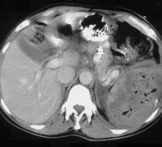
This picture that shows tiny bubbles in the spleen parenchyma that represent “normal” gas after embolization:

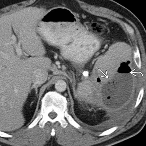
And the following one shows an air/fluid collection in the spleen that indicates an abscess:

Bottom line: Tiny bubbles in the spleen (and probably the liver) occur normally after angioembolization. They usually develop within an area of infarction, and most are benign. It is possible for them to evolve into a splenic abscess, but unlikely. Many embolization patients develop fevers at some point, and most have an elevated WBC count. So in most cases, you can ignore this incidental finding, as long as your patient has mild symptoms.

However, if the patient develops high fevers, very elevated WBC (> 25K), increasing abdominal or flank pain, and the spleen develops an air/fluid level, an abscess is forming. Despite what your radiologist might suggest, catheter drainage is not a good idea. The tubes are too small to remove the slurry that is generally found within the abscess. A trip to the OR is the only effective treatment, and splenectomy is generally the only option.

Related posts:

Reference: CT Findings after Embolization for Blunt Splenic Trauma. J Vasc Intervent Radiol 12(2):209-214, 2001.

Retained Hemothorax Part 3: VATS

I’ve written about the use of lytics to treat retained hemothorax over the past few days. Although it sounds like a good idea, we just don’t know that it works very well. And they certainly don’t work fast. Lengths of stay were on the order of two weeks in both studies reviewed.

The alternative is video assisted thoracoscopic surgery (VATS). So let’s take a look at what we know about it. This procedure is basically laparoscopy of the chest. A camera is inserted, and other ports are added to allow insertion of instruments to suck, peel, and scrape out the hemothorax.

A prospective, multi-center study was performed over a 2 year period starting in 2009. Twenty centers participated, contributing data on 328 patients with retained hemothorax. This was defined as CT confirmation of retained blood and clot after chest tube placement, with evidence of pleural thickening.

Here are the factoids:

  • 41% of patients had antibiotics given for chest tube placement (this is interesting given the lack of consensus regarding their effectiveness!)
  • A third of patients were initially managed with observation, and most of them (82%) did not need any further procedures (83 of 101 patients)
  • Observation was more successful in patients who were older, had smaller hemothoraces (<300cc), smaller chest tubes (!!, <34 Fr), blunt trauma, and peri-procedure antibiotics (?)
  • An additional chest tube was inserted in 19% of patients, image guided drain placement in 5%, and lytics in 5%. Half to two-thirds of these patients required additional management.
  • VATS was used in 34% of patients. One third of them required additional management including another chest tube, another VATS, or even thoracotomy.
  • Thoracotomy was most likely required if there was a diaphragm injury or large hemothorax (<900cc)
  • Empyema and pneumonia were common (27% and 20%, respectively)

Bottom line: There’s a lot of data in this paper. Most notably, many patients resolve their hemothorax without any additional management. But if they don’t, additional tubes, guided drain placement, and lytics work only a third of the time and contribute to additional time in the hospital. Even VATS and thoracotomy require additional maneuvers 20-30% of the time. And infectious complications are common. This is a tough problem!

Tomorrow, I’ll try to roll it all together and suggest an algorithm to try to optimize both outcomes and cost.

Posts in this series:

Reference: Management of post-traumatic retained hemothorax: A prospective, observational, multicenter AAST study. 72(1):11-24, 2012.

Retained Hemothorax Part 2: Lytics (again)

Yesterday, I reviewed a small case report that was published a couple of years ago on lytics for treatment of retained hemothorax. But surely, there must be something better, right?

After digging around, I did find a paper from 2007 that prospectively looked at protocolized management of retained hemothorax, and its aftermath. It was carried out at a busy Level I trauma center over a 16 month period.

All patients with a hemothorax treated with chest tube received daily chest x-rays. Those with significant opacification on day 3 underwent CT scan of the chest. If more than 300 cc of retained blood was present, the patient received streptokinase or urokinase (surgeon preference and drug availability) daily, and rolled around in bed for 4 hours to attempt to distribute it. The chest tube was then unclamped and allowed to drain. This was repeated for 3 days, and if there was still opacification, a repeat CT was obtained. If the volume was still greater than 300 cc, the cycle was repeated for the next 3 days. If the opacification cleared at any point, or the repeat CT showed less than 300 cc, the protocol was stopped and the chest tube removed. If the chest was still opacified after 6 days, VATS was offered.

Here are the factoids:

  • A total of 203 patients with hemothorax were admitted during the study period and 25 (12%) developed a retained hemothorax
  • While a few had treatment start within 4 days, the majority did not receive lytics until day 9 (range 3  –30 days!)
  • The average length of time in hospital after start of lytics was 7 days, leading to a total length of stay of 18 days
  • 92% of patients had “effective” evacuation of their retained hemothorax, although 1 had VATS anyway which found only 100 cc of fluid
  • 16 patients had “complete” evacuation, and 5 had “partial” evacuation
  • There were no hemorrhagic complications, but one third of patients reported significant pain with drug administration

Bottom line: Sounds good, right? The drug seems reasonably effective, although lengths of stay are relatively long. However, streptokinase and urokinase are no longer available in the US, having been replaced with tissue plasminogen activator (tPA). This paper does a cost analysis of lytics vs VATS and found that the former treatment cost about $15000 (drug + hospital stay) vs $34000 for VATS. However, a big part of this was that the drug only cost about $75 per dose. tPA is much more expensive.

So once again, small series, longer lengths of stay, but at least nicely done. Unfortunately, the drug choice is no longer available so use of tPA tilts the balance away from lytics. Tomorrow, I’ll explore the results of using VATS for this condition.

Posts in this series:

Reference: Intrapleural Thrombolysis for the Management of Undrained Traumatic Hemothorax: A Prospective Observational Study. J Trauma 62(5):1175-1179, 2007.

Retained Hemothorax Part 1: Lytics

Hemothorax is a common complication of chest trauma, occurring in about one third of cases. It is commonly treated with a chest tube, which usually takes care of the problem. But in a few cases some blood remains, which can result in an entrapped lung or empyema.

There are several management options. Historically, these patients underwent thoracotomy to peel out the fibrinous collection stuck to lung and chest wall. This has given way to the more humane VATS procedure (video assisted thoracoscopic surgery) which accomplishes the same thing using a scope. In some cases, another tube can be inserted, sometimes under CT guidance, to try to drain the blood.

So what about lytics? It’s fibrin, right? So why not just dissolve it with tissue plasminogen activator (tPA)? There have been very few studies published over the years. The most recent was in 2014. I’ll review it today, and another tomorrow. Finally, I’ll give you my thoughts on the best way to deal with retained hemothorax.

Here are the factoids:

  • This was a single center, retrospective review of data from 1.5 years beginning in 2009
  • A total of seven patients were identified, and most had hemothorax due to rib fractures. Three presented immediately after their injury, 4 were delayed.
  • Median time from injury to chest tube placement was 11 days
  • Median time the chest tube was in place was 13 days, with an average hospital stay of 14 days
  • Patients received 1 to 5 treatments, averaging 24mg per dose
  • There was one death in the group, unrelated to TPA treatment
  • No patient “required” VATS, but one underwent thoracotomy, which turned out to be for a malignancy

Bottom line: The authors conclude that tPA use for busting retained hemothorax is both safe and effective. Really? With only seven patients? The biggest problem with this study is that it uses old, retrospective data. We have no idea why these patients were selected for tPA in this 5-year old cohort of patients. Why did it take so long to put in chest tubes? Why did the chest tubes stay in so long? Maybe this is why they were in the hospital so long?

Plus, tPA is expensive. A 100mg vial runs about $6000. Does repeatedly using an expensive drug and keeping a patient in the hospital an extra week or so make financial sense? So it better work damn well, and this small series doesn’t demonstrate that.

Tomorrow, I’ll look at the next most recent paper on the topic, from way back in 2004.

Posts in this series:

Reference: Evaluation of chest tube administration of tissue plasminogen activator to treat retained hemothorax. Am J Surg 267(6):960-963, 2014.

Geriatric Week 5: Falls In The Elderly: The Consequences

Falls among the elderly are a huge problem. Our trauma service typically has 6-12 elders who have sustained significant injuries on it at any given time. About a third of people living at home over the age of 65 fall in a given year. At 80 years and up, half fall every year.

Because of this, falls are the leading cause of ED visits due to an injury for those over 65. What exactly are the societal consequences of all these falls? A yet to be published study from the Netherlands looked at injuries, costs and quality of life after falls in the elderly.

The top 5 most common injuries included simple wounds, wrist and hip fractures, and brain injuries. Although hip fracture typically was #5 in the 65-74 age groups, it was uniformly #1 in the 85+ group. Patterns were similar in both men and women. Interestingly, hip fractures were by far the most expensive, making up 43% of the cost of all injuries (total €200M). The next closest injuries by total cost, superficial injuries and femur fracture, made up only 7% of the total each!

As you can imagine, quality of life suffered after falls as well. A utility score based on the EQ-5D, a validated quality of life score, was lower in fall victims. Even after 9 months, this score did not return to baseline. About 70% of elders who were admitted after their falls described mobility problems and 64% had problems with their usual activities. Over a quarter expressed problems with anxiety or depression.

Bottom line: An array of falls prevention programs are available. They need to be more aggressively implemented to reduce costs and improve the quality of life of our elders.

Reference: Social consequences of falls in the older population: injuries, healthcare costs, and long-term reduced quality of life.  J Trauma 71(3):748-753, 2011.