Category Archives: Abdomen

What Would You Do? Teensy Weensy Stab To The Abdomen – Part 2

Yesterday, I presented the case of a young man with a teensy weensy little stab to his abdomen, just above the umbilicus. There was a tiny bit of oddly colored fat that was visible in the wound. So now what should we do?

The first thing is to figure out what that bit of fat is. It doesn’t have the normal large pebbling and color of subcutaneous fat. Therefore, it must be a small piece of omentum protruding from the wound.

And what is the significance of that? This question has been addressed by papers with low numbers of subjects since the 1980s. It really depends on what country you are located in. Do you have readily available OR resources? Are there pressures to minimize hospital stays (US)?

One of the earliest papers originated from Parkland Hospital in Dallas TX. They reviewed 115 cases of omental evisceration over a 4 year period, and found that “serious” abdominal injuries were found in 75% of them. All went to laparotomy, and injuries to not one, but two organs were noted in about half of the positive cases. There was a 7% complication rate with negative laparotomy,

Contrast this with a study from Kingston, Jamaica where 66 patients with abdominal stabs and omental evisceration were treated. Of these, 14 were treated with observation because they had a normal abdominal exam. All were treated successfully without operation. But note the ratio here: 14/66 = 21%, which is the same as the negative laparotomy in the Parkland study (25%). So this study implies that if the patient can be watched and does not develop symptoms, the negative lap may be avoided.

Unfortunately, in many countries there are pressures to get people out of the hospital as soon as possible. Since small bowel content is relatively benign (at first), patients may not become symptomatic for several days. It would probably be difficult to convince your hospital to keep patients laying around for serial exams for days on end. Not to mention the logistical problems of doing good serial exams.

So most trauma professionals will be compelled to do something. And what should we do? Here are some possibilities. Pick your poison, and I’ll give you my choice tomorrow.

  • Local wound exploration
  • CT scan of the abdomen
  • Proceed to the operating room

As before, leave a comment to let me know what you would do. Or tweet it out!

References:

  1. Significance of omental evisceration in abdominal stab wounds. Am J Surg 152(6):670-673, 1986.
  2. Non-operative management of stab wounds to the abdomen with omental evisceration. J Royal Col Surg Edin 41(4):239-240, 1996.
Print Friendly, PDF & Email

Best of AAST #10: Pediatric Contrast Extravasation And Pseudoaneurysms

There is a significant amount of variation in the management of pediatric solid organ injury. This is well documented between adult and pediatric trauma centers in t, but also apparently between centers in different countries. A poster from a Japanese group in Okinawa Japan will be presented this week detailing the relationship between contrast extravasation after spleen or liver injury and pseudoaneurysm formation.

In adults, the general rule is that pseudoaneurysms just about anywhere slowly enlarge and eventually rupture. This group sought to define this relationship in the pediatric age group. They performed a multi-center observational study of retrospectively enrolled children, defined as age 16 and less. Those who had contrast extravasation on initial CT were monitored for later pseudoaneurysm formation.

Here are the factoids:

  • 236 patients were enrolled across 10 participating centers, with about two-thirds having liver injury and the remainder with splenic injury
  • 80% of patients underwent followup CT scan (!!)
  • 33 patients (15%) underwent angiography (!!!!)
  • 17 patients with CT scan (2%) had pseudoaneurysm formation and 4 of them had a delayed rupture
  • Overall, pseudoaneurysms occurred in 29% of those with contrast extravasation and 5% without extravasation
  • The authors concluded that contrast extravasation was significantly associated with pseudoaneurysm formation after adjusting for variables such as ISS, injury grade, and degree of hemoperitoneum

Bottom line: This is an abstract, so a lot is missing. What was the age distribution, especially among those who underwent angiography? Was the data skewed by a predominantly teenage population, whose organs behave more like adults? The abstract answers a question but ignores the clinical significance.

For those trauma professionals who routinely care for pediatric patients, you know that contrast extravasation in children doesn’t act like its adult counterpart. Kids seldom decompensate, and for those who are mistakenly taken for angiography, the extravasation is frequently gone. The authors even admitted in the conclusion that aggressive screening and treatment for pseudoaneurysm was carried out.

The real question is, what is the significance of a solid organ pseudoaneurysm in children? Based on my clinical experience and reading of the US literature, not much. Of course, there is a gray zone as children move into adulthood in the early to mid-teens. But this does not warrant re-scanning and there should be no routine angiography in this age group. Contrast extravasation in pediatric patients warrants close observation for a period of time. But intervention should only be considered in those who behave clinically like they have ongoing bleeding. 

Reference: Association between contrast extravasation on CT scan and pseudoaneurysm in pediatric blunt splenic and hepatic injury: a multi-institutional observational study. Poster 31, AAST 2018.

Print Friendly, PDF & Email

The Handoff In Damage Control Surgery

Damage control is over 25 years old already! We continue to refine the techniques and closure techniques/devices, and have developed novel ways to speed closure of the abdominal wall in order to avoid pesky hernias. But the process itself is time intensive, and typically several days pass with regular returns to OR until closure is achieved.  This is one of the prime areas in which human error can occur, especially with modern service-style coverage of trauma patients.

In the old days, trauma patients were admitted by their surgeon, and that person provided their care nearly continuously until discharge. He or she rounded on them daily, took them back to the OR when needed, and then discharged them.

This is less practical (and desirable) in this day and age. And even if it seems possible, it’s not. No one can be on call 24 hours a day, and provide comprehensive care to every patient, around the clock. Many trauma programs have adopted a “service model”, where patients are admitted to a defined care team and managed by them. The team is led by a surgeon, but that person may change on a weekly (or in some cases nearly daily) basis. I call this the “interchangeable head” model, and to make it work there must be excellent handoffs during any leadership change.

In some cases, a patient may undergo a damage control procedure by one surgeon, but another must do the takeback and possibly the definitive closure. In this case, the handoff is critical! It is paramount that the next surgeon know everything about the first case so that they can perform the correct procedure.

How can this be accomplished? Here are some tips:

  • Do not rely on the medical record and previous operative note. It may not be available, and there is usually some loss of information in recording it anyway. Don’t believe it.
  • Ideally, meet face to face with the previous surgeon(s). Get the blow by blow description of exactly everything that was done and how. Also discuss what still needs to be done, and when. Try to maintain a uniform philosophy of patient care across surgeons.
  • If face to face is not possible, a telephone call is acceptable. The discussion is exactly the same.
  • If the surgery occurred at an outside hospital and was then transferred, you must call the initial surgeon to have this discussion before going to the OR!
  • If something unexpected is encountered during the case, make sure you have contact information so you can call during the case.

Applying these concepts will decrease the possibility of error, as well as the likelihood of any iatrogenic harm to these complex patients.

Print Friendly, PDF & Email

Best of: Blunt Duodenal Injury In Children

Blunt injury to hollow organs is rare in adults, but a little more common in children. This is due to their smaller muscle mass and the lack of protection by their more flexible skeleton. Duodenal injury is very rare, and most trauma professionals don’t see any during their career. As with many pediatric injuries, there has been a move toward nonoperative management in selected cases, and duodenal injury is no exception.

What we really need to know is, which child needs prompt operative treatment, and which ones can be treated without it? Children’s Hospital of Boston did a multicenter study of pediatric patients who underwent operation for their injury to try to tease out some answers about who needs surgery and what the consequences were.

A total of 16 children’s hospitals participated in this 4 ½ year study. Only 54 children had a duodenal injury, proven either by operation or autopsy. Some key points identified were:

  • The injury was very uncommon, with one child per hospital per year at best
  • 90% had tenderness or marks of some sort on their abdomen (seatbelt sign, handlebar mark, other contusions).
  • Free air was not universal. Plain abdominal xray showed free air in 36% of cases, while CT showed it only 50% of the time. Free fluid was seen on CT in 100% of cases.
  • Contrast extravasation was uncommon, seen in 18% of patients.
  • Solid organ injuries were relatively common
  • Amylase was frequently elevated

Although laparoscopic exploration was attempted in about 12% of patients, it was universally converted to an open procedure when the injury was confirmed. TPN was used commonly in the postop period. Postop ileus was very common, but serious complications were rare (wound infection <10%, abscess 3%, fistula 4%). There were 2 deaths: one child presented in extremis, the other deteriorated one day after delayed recognition of the injury.

Bottom line: Be alert for this rare injury in children. Marks on the abdomen, particularly the epigastrium, should raise suspicion of a duodenal injury. The best imaging technique is the abdominal CT scan. Contrast is generally not helpful and not tolerated well by children. Duodenal hematoma can be managed nonoperatively. But any evidence of perforation (free fluid, air bubbles in the retroperitoneum, duodenal wall thickening, elevated serum amylase) should send the child to the OR. And laparotomy, not laparoscopy, is the way to go.

Reference: Operative blunt duodenal injury in children: a multi-institutional review. J Ped Surg 47(10):1833-1836, 2012.

Print Friendly, PDF & Email

Off-Label Foley Use In Trauma – Part 2

Yesterday, I wrote about an unusual way to use the Foley urinary catheter to plug a heart wound. This allows you to buy time to get to the operating room to perform the definitive repair. But this cheap and effective tool is very versatile, and can be used in other body areas as well.

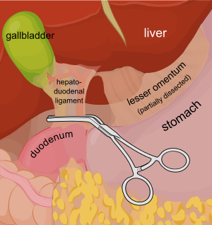
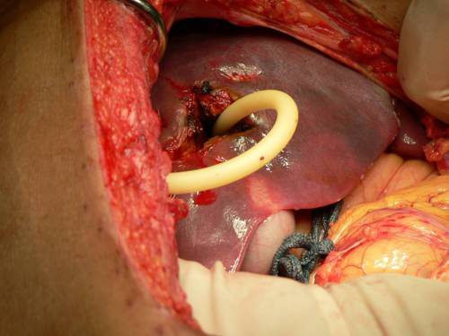
Consider a deep penetrating injury to the liver. It takes time to determine which method for slowing/stopping the bleeding is most appropriate. Sure, the doctor books say to occlude the inflow by gently clamping the hepatoduodenal ligament (Pringle maneuver). But this takes time, and can be difficult if there is lots of bleeding.

You may be able to gain some time by placing a properly sized Foley catheter directly into the wound and carefully inflating with saline. You must inflate the balloon to feel, not to its full volume. It should be snug, but not so full that it cracks the liver parenchyma and causes yet more bleeding.

Bottom line: Any time you find yourself facing bleeding from hard to expose places, think about using a balloon catheter like the Foley. Sizing is critical, and the balloon volume is more important than the catheter diameter. Estimate the size of the area that needs to be occluded, and then ask for a catheter with a 10cc or 30cc balloon. If you need smaller, more precise control, try a Fogarty arterial embolectomy catheter instead. 

As with the cardiac Foley, be sure to occlude the end so you don’t create a conduit for the blood to escape. If your patient does well, and you need to leave the catheter in place for a damage control closure, LEAVE THE CATHETER COMPLETELY WITHIN THE ABDOMEN. If you exteriorize the end, some well-meaning person may unclamp it, drop the balloon, or decide that it can be used for tube feedings.

TIP: If the distance between the balloon and the catheter tip is too long, DO NOT TRY TO SHORTEN THE TIP BY CUTTING IT! This will damage the balloon and it will not inflate.

Fogarty catheters

Print Friendly, PDF & Email