Category Archives: Abdomen

AAST 2019 #3: Delayed Splenectomy In Pediatric Splenic Injury

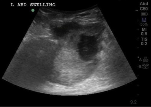
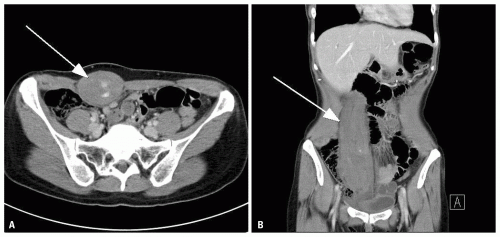
Nonoperative management of the blunt injured spleen is now routine in patients who are hemodynamically and have no evidence of other significant intra-abdominal injury.  The trauma group at the University of Arizona – Tucson scrutinized the failure rate of this procedure in children because it is not yet well established.

They reviewed 5 years of data from the National Readmission Database. This is actually a collection of software and databases maintained by the federal government that seeks to provide information on a difficult to track patient group: those readmitted to hospitals after their initial event.

Patients who had sustained an isolated spleen injury who were less than 18 years old and who had either nonoperative management (NOM), angioembolization (AE), or splenectomy were analyzed. Outcome measures included readmission rate, blood transfusion, and delayed splenectomy. Common statistical techniques were used to analyze the data.

Here are the factoids:

  • About 9500 patients were included, with an average age of 14
  • Most (77%) underwent NOM, 16% had splenectomy, and 7% had AE (no combo therapies?)
  • Significantly more patients with high grade injury (4-5) had splenectomy or AE than did the NOM patients (as would be expected)
  • A total of 6% of patients were readmitted within 6 months of their initial injury: 12% of NOM *, 8% of AE *, and 5% of those with splenectomy (* = statistically significant)
  • The NOM and AE patients were also more likely to receive blood transfusions during their first admission
  • Delayed splenectomy occurred in 15% of cases (7% NOM and 5% AE) (these numbers don’t add up, see below)
  • Statistical analysis showed that delayed splenectomy was predicted by high grade injury (of course), blood transfusion (yes), and nonoperative management (huh?)
  • In patients who were readmitted and splenectomized, it occurred after an average of 14 days for the NOM group and 58 days for AE (huh?)

The authors concluded that “one in seven children had failure of conservative management and underwent delayed splenectomy within 6 months of discharge.” They stated that NOM and AE demonstrated only a temporary benefit and that we need to be better about selecting patients for nonoperative management.

Hmm, there are several loose ends here. First, what is the quality of the study group? Was it possible to determine if these patients had been treated in a trauma center? A pediatric vs adult trauma center? We know that there are outcome disparities in spleen trauma care at different types of trauma centers. 

Next, are they really pediatric patients? Probably not, since age < 18 were included and the average age was 14. Injured spleens in pre-pubescent children behave much better than adolescents, which are more adult-like.

And what about the inherent bias in the “readmission data set?” You are looking only at patients who were readmitted! By definition, youare looking at a dataset of poorer outcomes. What if you had identified 9,500 initial patient admissions from trauma registries and then tried to find them in the readmission set. I know it’s not possible to do that, but if it were I would bet the readmission and delayed splenectomy numbers would be far, far lower.

And what about those delayed splenectomy numbers? I can’t get the percentages to match up. If 15% of the 7965 patients who didn’t have an initial splenectomy  had it done later, how does 7.2% of the 7318 NOM patients and 5.3% of the 1541 AE patients add up?

Bottom line: The usual success rate tossed around for well-selected nonoperative management is around 93% when optional adjunctive AE is part of the algorithm. That’s a 1 in 14 failure rate, and it generally occurs during the initial hospitalization. In my experience, readmissions are very rare. And that’s for adults; children tend to behave even better!

I wouldn’t consider changing my practice yet based on these findings, but the devil will probably be in the details!

Here are some questions for the presenter and authors:

  • Please provide some detail on the data set. We really need to know an age breakdown and the types of centers they were treated at, if available.
  • Discuss the potential data set bias working backwards from a database that includes only readmitted patients.
  • Please clarify the delayed splenectomy statistics to help match up the numbers.

I’m anticipating a great presentation at the meeting!

Reference: Delayed splenectomy in pediatric splenic injuries: is conservative management overused? AAST 2019 Oral abstract #8.

Print Friendly, PDF & Email

How To: Manage Rectus Sheath Hematomas

Although not strictly traumatic, rectus sheath hematomas frequently come to the attention of trauma professionals. In some cases, they may be due to vigorous physical activity or blunt impact. They may also occur spontaneously, especially in patients who are anticoagulated.

This is not a very common condition, accounting for only 1-2% of patients who present with acute abdominal pain.  The common etiology of rectus hematomas is either a tear of the major blood supply (superior and inferior epigastric arteries) or a tear of the muscle itself with bleeding from smaller vessels. The loose attachment of the inferior epigastric  and the fixed perforating  muscular branches make injury in the lower half of the muscle more common.

These hematomas are frequently self-limiting problems. The rectus sheath provides containment for the hematoma, and as pressure rises, bleeding slows and stops. However, if the hematoma is able to escape posteriorly, it can result in life-threatening bleeding.

Presentation generally consists of abrupt onset of focal abdominal pain, and an abdominal wall mass. The pain can be rather intense, making it difficult to determine if it is intraperitoneal or in the body wall. Tip: ask the patient to tense their abdominal wall muscles, then palpate the area. If the tenderness increases, then it is more likely due to an abdominal wall source. Tensing the muscles will shield sources inside the peritoneal cavity, decreasing tenderness to palpation.

Diagnosis may be made by physical exam, but not always. The hematoma may be seen using ultrasound, but the gold standard is the contrast-enhanced CT. Contrast is essential to determine if active extravasation is occurring.

Ultrasound

CT scan with contrast showing extravasation

Patients who are hemodynamically stable and do not have active extravasation may be treated conservatively. However, a significant number of patients will require at least one unit of blood. Be prepared and send a type and crossmatch. Conservative management includes ice packs for pain relief, direct pressure (sand bags), and reversal of anticoagulation if possible. Stable patients with extravasation on CT should be evaluated by angiography and embolized if a bleeding vessel can be identified.

Unstable patients must be resuscitated promptly with fluid and blood so they can be taken to the angiography suite. Operative exploration is extremely unsatisfying and should be avoided, since it is difficult to find the bleeding vessels in the midst of a huge hematoma. 

Print Friendly, PDF & Email

Stab To The Abdomen: The WTA Algorithm

I’ve spent the last week discussing the hypothetical case of a young patient with a stab to the abdomen. I worked through some of the thought processes regarding physical exam, imaging, and choices for management. Fortuitously, it would seem, The Journal of Trauma published an algorithm on this very topic from the Western Trauma Association (WTA).

The WTA Algorithm Committee reviewed existing data to start the process of developing this algorithm. As could be expected, very little high quality data was available. So the final algorithm is a synthesis of existing lesser quality studies, expert opinion from the committee members, and commentary from the membership.

Here are some of the highlights:

  • Unstable patients go straight to the operating room (A)
  • Patients who cannot be examined (unconscious, head injured, intoxicated) should be evaluated for peritoneal penetration with local wound exploration, ultrasound, CT, or laparoscopy. If positive or equivocal, proceed to exploration. (B)
  • Patients who can be examined should be managed by location of the stab. Flank injuries are lower risk and should be scanned. Anterior stabs can be evaluated using observation, local would exploration, or CT scan,
  • Positive results generally proceed to laparotomy. The algorithm states that laparaoscopy “may be performed in select stable patients by a highly skilled surgeon experienced in minimally invasive surgical techniques.”

As with any algorithm or practice guideline, nothing is etched in stone. These tools are good for about 90% of the clinical situations you will encounter. If you end up off the beaten path, you will need to use your best judgment to provide best treatment for your patient. Just remember to document your rationale, because you may very well have to justify it to your peers.

Click the diagram below to see a full size version.

Reference: Evaluation and management of abdominal stab wounds:
A Western Trauma Association critical decisions algorithm. J Trauma 85(5):1007-1015, 2018.

Print Friendly, PDF & Email

What Would You Do? A Teensy Weensy Stab To The Abdomen – Part 4

We’ve gotten the young man with the teensy weensy stab to the abdomen with a bit of omental evisceration to the operating room. Now what should we do? We’ve already decided that he needs an exploration because of the known penetration. How should we go about it?

There are two choices: diagnostic laparoscopy vs laparotomy. Which is better? Let’s talk about laparoscopy first. This tool has been around now for over 25 years. There has been variable acceptance for use in trauma during that time because it tends to take more time and may have a higher rate of missed injury. Both factors have major implications in patients who have active bleeding and small injuries, respectively.

On the plus side, a truly negative (nontherapeutic) exploration tends to be more benign, with rapid recovery, faster time to discharge, and potentially fewer complications when evaluated with a scope. But on the minus side, small injuries can be notoriously difficult to find. What does that small wisp of blood mean? This is not nearly as clear as the meaning of other colors (green, brown). The decision to open can be difficult, particularly for surgeons who perform a high number of laparoscopies in the non-trauma portion of their practice.

Trauma laparotomy is traditionally a large operation with a generous incision and meticulous exploration. This can lead to significant postop pain and morbidity, particularly when no significant pathology is found. Unfortunately, the literature appears to be quite polarized. The surgeon is either pro-laparoscopy, or pro-big incision, and tends to brace their preferred procedure almost exclusively.

But there is a middle ground, and that is what I would choose in a case like this. The surgeon must consider the likelihood of reliably finding the size of internal injury based on his or her assessment of the external wound, as well as the probability that the exploration would be non-therapeutic. So in this case, I would worry that a bowel injury could be only a few millimeters in size and might be missed using only the laparoscope. But I also think that there is a good chance there may not be an injury at all, so I would not be inclined to start with a huge incision.

My choice is to perform a “mini-laparotomy”, making an incision just large enough to explore all of the bowel and visualize the retroperitoneum. I can generally do this through an incision large enough to get my palm into the abdomen, about 6cm. I am confident that I can easily find all injuries, and make the incision larger if warranted. Postoperative pain is better, and discharge if no injuries were found can happen in 1-2 days.

Unfortunately, I can’t find any papers that examine this middle ground between laparoscopy and full laparotomy. But I’ll keep looking! How would you have managed this case? Comment or tweet, please!

In my next post, I’ll review the official algorithm for evaluating stabs to the abdomen recently published by the western Trauma Trauma Association.

References: 

  1. The role of laparoscopy in management of stable patients with
    penetrating abdominal trauma and organ evisceration. J Trauma 81(2):307-311, 2016.
  2. Diagnostic Laparoscopy for Trauma: How Not to Miss Injuries. J Laparoscopic Adv Surg Tech 28(5):506-513, 2018.
Print Friendly, PDF & Email

What Would You Do? Teensy Weensy Stab To The Abdomen – Part 3

In my last post, I described the plight of a young man who had sustained a stab to the abdomen. It appeared that a very tiny bit of omentum was hanging out of the wound. What to do?

I listed three options:

  • Local wound exploration
  • CT scan of the abdomen
  • Proceed to the operating room

So let’s work through these. First, local wound exploration (LWE).

LWE is a diagnostic procedure to determine if a sharp wound has actually or potentially penetrated a vital area. It is usually performed in the neck to determine if the platysma has been violated, or in the abdomen top check for peritoneal violation. In this case, you would use it if you just couldn’t believe that the bit of odd fat was actually omentum, or if you were unsure what you were looking at. You could also grab it (gently) and give it a little tug. If more comes out, you’ve made your diagnosis. Fortunately, this is rarely necessary because omentum has a very distinctive appearance. You know it when you see it.

What about probing the wound? One of my mentors, John Weigelt, used to ask, “Michael, does your finger / q-tip / instrument have an eyeball on the end of it?” His point was that probing is like so many other medical tests: diagnostic if positive, but unsettling if it’s not. What happens if the wound does penetrate, but you can’t find the path that the knife/bullet took? You can only call that indeterminate. I suppose you could take an approach that includes probing first, then proceeding to full LWE if that is negative.

I’ll describe the proper technique for local wound exploration in a later post.

And what about CT scan? This is another unsatisfying test, because it is very likely to be negative with small wounds. The fascial defect in this case will be very small, and can easily be missed on the scan. Not recommended.

Given all this discussion, my vote is to proceed to the operating room. I know this is omentum, and I know that there is a good likelihood that there will be an injury that needs repair. So let’s go get it done.  But what procedure should I do, and how should I do it? That’s the subject for my next post.

As always, please leave comments below or tweet them out!


Print Friendly, PDF & Email