Here’s a short, 5 minute video on how to grade spleen injuries like a pro! Enjoy!

Related posts:
- Solid organ injury tips
- Platelet count after spleen injury
- Bedrest after pediatric solid organ injury? Really?

Here’s a short, 5 minute video on how to grade spleen injuries like a pro! Enjoy!
Related posts:
Most trauma centers have some kind of practice guideline for managing solid organ injury. Unfortunately, the specifics at each center are all over the map. Here are a few common questions:
As for activity, some earlier studies have shown that early ambulation is safe. The group at Hahnemann University Hospital in Philadelphia tried to determine if early mobilization would decrease time in ICU and/or the hospital, or increase complications.
Until 2011, their trauma service kept all patients with solid organ injury at bed rest for 3 days(!). They modified this routine to allow ambulation the following morning for Grade 1 and 2 injuries, and after 24 hours for Grade 3 and above, or those with hemoperitoneum. They examined their experience for 4 years prior (PRE) and 4 years after (POST) this change. They excluded patients with penetrating injury, or other significant injuries that would impact the length of stay.
Here are the factoids:
Bottom line: It’s about time we recognized what a waste of time these restrictions are! Unfortunately, the study groups became very small after exclusions, but apparently the statistics were still valid. But still, it continues to become clear that there is no magic in keeping someone starving in their bed for any period of time.
At my hospital, we adopted a practice guideline very similar to this one way back in 2004 (download it below). Hospital lengths of stay dropped to about 1.5 days for low grade injury, and to about 2.5 days for high grade.
And earlier this year, we eliminated the NPO and bed rest restrictions altogether! How many patients actually fail and end up going urgently to the OR? So why starve them all? And normal activity started immediately is no different than activity started a few hours or days later.
Don’t starve or hobble your patients, adults or children!
Related posts:
Reference: Early mobilization of patients with non-operative liver and spleen injuries is safe and cost effective. AAST 2016, Poster #5.
The shift toward initial nonoperative management of spleen injuries began in the early 1990’s, as the resolution of early CT scans began to improve. Our understanding of the indicators of failure also improved over time, and success rates rose and splenectomy rates fell.
Angiography was adopted as an adjunct to early management, especially when we figured out what contrast extravasation and pseudoaneurysms really meant (bad news, and nearly certain failure in adults). At first, it was used in a shotgun approach in most of the higher grade injuries. But we have refined it over the years, and now it is used far more selectively at most centers.
A group at Indiana University was interested in looking at the impact of angio use on splenic salvage over a long time frame. They queried the National Trauma Data Bank, looking specifically at high grade splenic injury care at Level I and II centers from 2008-2014. Patients undergoing splenectomy were divided into early (<= 6hr after admission) and late (> 6 hrs). Over 50,000 records were analyzed.
Here are the factoids:
So the authors recognize that late splenectomy has decreased. But they also state that early splenectomy has increased. They attribute it to increased recognition of patient requiring early splenectomy. They then call into question the need to use angiography if it hasn’t decreased the overall splenectomy rate.
Problem: The early splenectomy rate increased from about 13% to 14%, reading their graph, and is probably not significant. These are the failures that occur in the trauma bay and shortly thereafter that must be taken to the OR. The late splenectomy rate decreased from 5% to 3%, which may be significant (p value not included in the abstract). These are failures during nonoperative management, and are decreasing over time. And BTW, the authors do not define what “high grade” splenic injuries they are looking at.
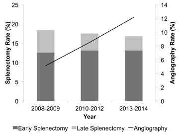
Bottom line: This abstract illustrates why it is important to read the entire article, or in this case, listen to the full presentation at AAST. It sounds like one that’s been written to justify not having angiography available as it is currently required.
The authors showed that overall splenectomy rate was the same, but delayed splenectomy (late failure) has decreased with increasing use of angiography. But remember, this is an association, not cause and effect. Most of the early failures are still probably ones that can’t be prevented, but we’ll see if the authors can dissect out how many went to OR very early (not eligible for angio), or later in the 6 hour period (could have used angio). It looks to me like the use of angiography is having the desired effect. But undoubtedly we could use that resource more wisely. What we really need are some guidelines as to exactly when a call to the interventional radiologists is warranted.
Related posts:
Reference: Overall splenectomy rates remain the same despite increasing usage of angiography in the management of high grade blunt splenic injury. AAST 2016, paper 35.
Over the years, I’ve written about solid organ injury management many times. Here is a summary of some practical pointers and tips, some old and some new. They are as evidence-based as I can get them. This kind of stuff is not always in the doctor and nursing books.
After discharge, our usual orders are:
Related post:
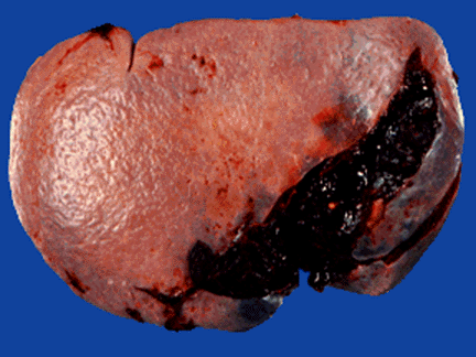
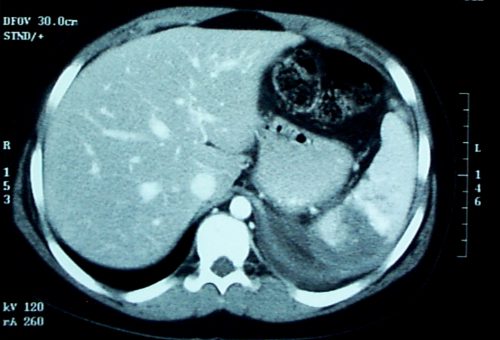
Contrast blush is always a concern when seen on CT of the abdomen for trauma. It can represent one of two things, and both are bad:
These two clinical issues can be distinguished by looking at the location of the contrast and its persistence. A pseudoaneurysm is located within the parenchyma, and the contrast will wash away, so it will not be visible on delayed images. Contrast that extends beyond the parenchyma or persists in delayed views represents active bleeding. In either case, the failure rate of nonoperative management exceeds 80% in adults without additional measures being taken.
Clinically, these patients usually act as if they are losing volume and require additional crystalloid and/or blood transfusion. The natural history in adults is for bleeding to continue or for the pseudoaneurysm to rupture, resulting in a quick trip to the operating room.
If vital signs can be maintained with fluids and blood, a trip to interventional radiology may solve the problem. Selective or nonselective embolization can be carried out and patients with only a few bleeding points can be spared operation. However, if multiple bleeding areas are seen, it is probably better to head to the OR for splenorrhaphy or splenectomy.
The image below shows likely areas of extravasation. They are a bit large to be pseudoaneurysms.
Children are different than adults. Extravasation from spleen injuries in prepubescent children frequently stops on its own. Angiography should only be used if the child is failing nonoperative management.