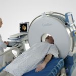
Blunt Trauma Radiographic Imaging Protocol

Last year, we developed an evidence-based protocol for deciding what radiographic images to order in our blunt trauma patients. For some body regions, there is fairly good literature available for guidance (i.e. Canadian head and cervical spine rules). For other areas, there is not nearly as much.

We convened a small group of people, including trauma surgeons, emergency physicians, radiologists and a radiation physicist to combine the information into a practical tool. 

You can view or download the worksheet we use by clicking the link at the bottom of this post. The protocol has been in use for about 9 months, and has significantly decreased the use of higher radiation dose imaging (CT). As a result, there has been a small increase in the use of lower dose conventional imaging (plain spine studies), but no missed injuries. 

Tomorrow, I’ll write about the specifics of how this protocol has changed our ordering habits. Click here to view it.

Click here to download the Blunt Trauma Radiographic Imaging Protocol Worksheet

Click here to download a bibliography of the literature used to develop the protocol

Double CT Scanning in Trauma

Typical order: “chest CT with and without contrast”

A review of Medicare claims from 2008 showed that 5.4% of patients received double CT scans of the chest. Although the median was about 2% across 3,094 hospitals, 618 hospitals performed double scans on more than 10% of their patients. And 94 did it on more that half! One of the outliers was a small hospital in Michigan that double scanned 89% of Medicare patients! As expected, there was wide variation from hospital to hospital, and from region to region around the US. 

Time for some editorial comment.

This practice is very outdated and shows a lack of understanding of the information provided by CT. Furthermore, it demonstrates a lack of concern for radiation exposure by both the ordering physician and the radiologist, who should know better. 

Some officials at hospitals that had high scan rates related that radiologists ordered or okayed the extra scan because they believed that “more information was better.” There are two problems with this thinking.

  1. Information for information’s sake is worthless. It is only important if it changes decision making and ultimately makes a difference in outcome. 
  2. As with every test we do, there may be false positives. But we don’t know they are false, so we investigate with other tests, most of which have known complications. 

The solution is to do only what is clinically necessary and safe. The tests ordered should be based on the best evidence available, which demands familiarity with current literature.

In trauma, there are a few instances where repeat scanning of an area is required. Examples include solid organ lesions which may represent an injury or a hemangioma, and CT cystogram to exclude bladder trauma. In both cases, only a selected area needs to be re-scanned, not the entire torso.

Bottom line: Physicians and hospitals need to take the lead and rapidly adopt or develop guidelines which are literature-based. State or national benchmarking is essential so that we do not continue to jeopardize our patient’s safety and drive up health care costs.

Tomorrow I’ll share the blunt trauma imaging protocol we use which has decreased trauma CT use significantly at Regions Hospital.

Related posts:

Trauma Mortality vs Cancer Mortality from CT Scans for Trauma

Trauma professionals worry about radiation exposure in our patients. A lot. There are a growing number of papers dealing with this topic in the journals every month. The risk of dying from cancer due to CT scanning is negligible compared to the risk from acute injuries in severely injured patients. However, it gets a bit fuzzier when you are looking at risk vs benefit in patients with less severe injuries. Is it possible to quantify this risk to help guide our use of CT scanning in trauma?

A nice paper from the Mayo clinic looked at their scan practices in 642 adult patients (age > 14) over a one year period. They developed dose estimates using a detailed algorithm, and combined them with data from the Biological Effects of Ionizing Radiation VII data. The risk level for injury was estimated using their trauma team activation criteria. High risk patients met their highest level activation criteria, and intermediate risk patients met their intermediate level activation criteria.

Key points in this article were:

  • Average radiation dose was fairly consistent across all age groups (~25mSv)
  • High ISS patients had a significantly higher dose
  • Cumulative risk of cancer death from CT radiation averaged 0.1%
  • This risk decreased with age. It was highest in young patients (< 20 yrs) at 0.2%, and decreased to 0.05% in the elderly (> 60 yrs)

Bottom line: Appropriate CT scan use in trauma evaluation is challenging. It’s use is widespread, and although it changes management it has not decreased trauma mortality. This paper shows that the risk of death from trauma in the elderly outweighs the risk of death from CT scan radiation. However, this gap narrows in younger patients with less serious injuries because of their very low mortality rates. Therefore, we need to focus our efforts to reduce radiation exposure on our young patients with minor injuries.

Related posts:

References:

  • Comparison of trauma mortality and estimated cancer mortality from computed tomography during initial evaluation of intermediate-risk trauma patients. J Trauma 70(6):1362-1365, 2011.
  • Health risks from low levels of ionizing Radiation: BEIR VII, Phase 2. Washington DC: The National Academies Press, 2006.

Extubation in the Emergency Department

Many patients are intubated in the emergency department who need brief control of their airway or behavior. In some cases, the condition requiring intubation resolves while they are still in the department. Most of the time these patients are admitted, typically to an ICU bed, for extubation. This is expensive and uses valuable resources. Is it possible to safely extubate these patients and possibly send them home?

Maryland Shock Trauma and Mount Sinai Medical Center looked at their experience in extubating selected patients in the ED. They looked at a series of 50 patients who were intubated for combativeness, sedation, or seizures. A specific protocol was followed to gauge whether or not extubation should be attempted.

None of the patients who were extubated per protocol required unplanned reintubation. One patient underwent planned reintubation when taken to the OR for an orthopedic procedure. 16% of patients were able to be discharged home from the ED.

Bottom line: A subset of patients who are intubated in the emergency department can be extubated once the inciting factor has resolved. These factors include sedation for painful procedures and combativeness. Following this protocol can reduce admission rates and reduce the use of scarce intensive care unit resources.

Click here to download a copy of the ED extubation protocol.

Related post: Trauma 20 years ago: ED intubation for head injury

Reference: Trauma patients can be safely extubated in the emergency department. J Emerg Med 40(2):235-239, 2011.

NOTE: The EMCrit blog, written by Scott Weingart, covered this topic last November. He is the first author on the paper and has created a nice podcast on the topic. You can find his blog here, and you can download the podcast here.

Early Coagulopathy After Severe Traumatic Brain Injury Equals Poor Outcome

Coagulopathy is a frequent occurrence after severe traumatic brain injury (TBI). There are high levels of tissue factor (TF) in the brain, which can be released with severe injury. This in turn triggers a cascade which can lead to generalized coagulopathy.

The trauma group at LAC+USC looked at the time course of coagulopathy after isolated severe TBI. They identified 278 patients over a 1.5 year period and retrospectively review a number of demographic and outcome variables. Coagulopathy was defined as a platelet count < 100,000/mm3, INR > 1.4, or PTT > 36 sec.

They found the following:

  • 46% with blunt trauma and 82% with penetrating injury developed a coagulopathy 
  • Presence of coagulopathy increased with increasing head injury severity
  • Thromobocytopenia as a cause of coagulopathy was less common (17%) than clotting factor problems
  • As brain injury severity increased from AIS=3 to AIS=5, median onset of coagulopathy became increasingly earlier (26 hrs, 22 hrs, 10 hrs)
  • Mortality increased with earlier coagulopathy (23% after 24 hrs, 39% between 12 and 24 hrs, 56% less than 12 hrs)

Bottom line:

  • Prehospital: Coagulopathy should be suspected if the patient is bleeding profusely from multiple sites, including your IV needle sticks. This indicates severe brain injury and demands triage to a trauma center with immediate neurosurgical support.
  • In-hospital: Coagulopathy that is noted in the ED portends severe injury and poor prognosis. Rapid access to CT scan and your neurosurgical consultant is critical.

Related post: Controlling fever in head injury

Reference: Time course of coagulopathy in isolated severe traumatic brain injury. Injury 41:924-928, 2010.

Home of the Trauma Professional's Blog

Do you want to get a daily email every time there’s a new post? See what I’m up to.

Click here to get details and subscribe!

[accua-form fid=”1″]

[mc4wp_form id=”2023″]