All posts by The Trauma Pro

Best Of AAST 2021: Reducing Errors In Trauma Care

Finally, a performance improvement (PI) abstract at AAST!

As many of you know, there are two general types of issues that are encountered in the usual PI processes: provider (peer) vs system. Provider issues are errors of omission or commission by an individual clinician. Examples include a surgeon making a technical error during a procedure, or prescribing the wrong drug or dose for some condition.

One might think that provider issues are the most common type of problem encountered. But they would be wrong. The vast majority of clinicians go to work each day with the idea that they will do their job to the best of their abilities. So how could things go awry?

Because the majority of errors have some degree of system component! They are set up to fail by factors outside their perception and/or control. Let’s look at a surgeon who has several small bowel anastomoses fall apart. His surgery department head chides/educates him, reports him to hospital quality, and proctors his next ten bowel cases. Everything is good, right?

But then, two months later, the stapler company issues a recall because they found a higher than usual number of anastomotic failures with one of their products. So it wasn’t the surgeon after all, like everyone assumed. This is an extreme example, but you get the idea. System issues often look like peer issues, but it’s frequently difficult for many PI programs to recognize or accept this.

A multi-institutional group reviewed the results of a newly implemented Mortality Reporting System (MRS) to analyze a large number of PI opportunities for improvement (OFI). More than 300 trauma centers submitted data to the MRS when a death occurred where an OFI was identified. The reports included details of the incident and mitigation strategies that were applied.


Here are the factoids:

  • A total of 395 deaths were reviewed over a two year period
  • One third of deaths were unanticipated (!!), and a third of those were failure to rescue
  • Half of errors pertained to clinical management, clinical performance, and communication
  • Human failures occurred in about two thirds of cases
  • The most common remedy applied was education, which presumes a “provider issue”
  • System strategies like automation, standardization, and fail-safe approaches were seldom used, implying that system issues were seldom recognized
  • in 7%, the trauma centers could not identify a specific strategy to prevent future harm (!!!)

The authors concluded that most strategies to reduce errors focus on individual performance and do not recognize the value of system-level intervention.

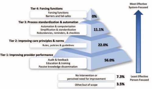
Bottom line: Look at the pyramid chart above (interesting choice for a chart, but very effective). The arrow shows progression from provider focus to systems focus. The pyramid shows how the recognition of and intervention for system issues drops off very rapidly.

I am both shocked and fascinated by the last bullet point. A strategy couldn’t be developed to prevent the same thing from happening again. Now, there are a few rare instances where this could be correct. Your patient could have been struck by a bolt of lightning in her room, or a meteorite could have crashed through the wall. But I doubt it. This 7% illustrates the importance of investigating all the angles to try to determine how the system failed!

For once, I have no critique for an abstract. It is a straightforward descriptive study that reveals an issue that many in PI are not fully aware of. I’ll definitely be listening to this one, and I really look forward to the published paper!

Reference: ERROR REDUCTION IN TRAUMA CARE: LESSONS FROM AN ANONYMIZED, NATIONAL, MULTI-CENTER MORTALITY REPORTING SYSTEM. AAST 2021, Oral abstract #17.

Best Of AAST 2021: Are We Getting Better At Balanced Resuscitation?

The way we resuscitate major trauma patients has been changing over the past decade. Even the 10th edition of the ATLS course has recognized that so-called balanced resuscitation is important. This concept limits the use of crystalloid and relies more heavily on blood component administration in ratios that more closely approximate whole blood.  Balanced resuscitation typically translates as the use of less than two liters of crystalloid, and blood product transfusion ratios of 1:1 to 2:1 (PRBC to plasma).

We have also recognized the critical importance of rapid control of major hemorrhage, which is best accomplished in an operating room.  The group at the University of Arizona massaged the TQIP database to see if these changes are having a significant impact on our patients.

They looked at five years worth of data, specifically reviewing information on adult patients with both transfusion and laparotomy occurring within four hours of arrival. The authors performed regression analyses to identify trends over the study period.

Here are the factoids:

  • Nearly 10,000 patients met study criteria with a mean age of 44 and ISS 34
  • Patients were in shock, with mean SBP 78 and median number of transfusions of 9 PRBC and 6 plasma
  • Time to laparotomy decreased from 1.87 hours to 1.37 hours over the five year period
  • 24-hour mortality decreased from 23% to 19% during the study
  • Blood product ratio decreased from 1.93:1 to 1.73:1
  • The authors state that the blood product ratio was independently associated with 24 hour mortality (odds ratio of 1.09) and in-hospital mortality (1.10) (??)

The authors conclude that resuscitation is becoming more balanced and time to surgery shorter, with a significant improvement in mortality.

Bottom line: Well, this is an interested study of associations. It uses a large database, which of course limits some of the information available. There are obvious trends toward faster time to OR (by 30 minutes) and a 4% improvement in survival. But the transfusion ratio really looks to be about the same. 

Let’s do the math, assuming that an average of 10 units of PRBC were given. A ratio of 1.93 would mean that 5.2 units of plasma were give (1425 cc, assuming 275 cc per unit). The ratio of 1.73 noted in 2017 would then be 5.8 units, or 1590 cc. This is an increase in FFP transfusion of 165 cc.

The authors stated that the improvement in transfusion ratios was statistically associated with the improvement in survival. I think this is one of those situations where there is a big difference between statistical significance and clinical significance. Do you really think that giving just 165 additional ccs of plasma could have that much overall effect on survival?

My suspicion is that there is a true association between the more rapid time to OR (and presumably surgical control) and survival. It’s just that the numbers were not clean enough to meet statistical rigor.

This is an interesting abstract, and shows that we are slowly getting better at controlling bleeding. But I think the most important takeaway is that we are not as good at balanced resuscitation as we think we are. We seem to be hovering at the 2:1 ratio, and only very slowly moving toward 1:1.

Questions for the authors / presenter:

  • Were you able to see a correlation between time to OR and survival?
  • Please comment on the association between transfusion ratios and survival, especially given the very small change over time.
  • Please clarify the in-hospital mortality and 24-hour mortality variables. In-hospital mortality suddenly pops up at the end of the results, but was never mentioned before.

Reference: AFTER 9,000 LAPAROTOMIES FOR BLUNT TRAUMA, RESUSCITATION IS BECOMING MORE BALANCED AND TIME TO INTERVENTION SHORTER: HOW LOW CAN WE GO? AAST 2021, Oral abstract #3.

Best Of AAST 2021: Hard Signs Of Vascular injury

Well, it’s that time of year again! The annual American Association for the Surgery (AAST) is just a few weeks away. Starting today, I will begin reviewing some of the interesting abstracts (to me, at least) that will be presented. I’ll give my analysis and perspective, and usually provide some questions for the presenters that they may face during the live meeting. Enjoy!

I’ll start with abstract #1. This one is from the AAST Prospective Observational Vascular Injury Trial (PROOVIT).  The group was established to create an aggregate database of information on the presentation, diagnosis, management (acute and definitive), surveillance and outcomes following vascular trauma. It manages a registry that collects a wide variety of data on assorted vascular injuries.

This study re-examines our use of “hard signs” to diagnose vascular injury. Back in the day, we had “hard signs” and “soft signs.”  Hard signs were fairly obvious indicators of serious injury, such as pulselessness, ischemia, pulsatile bleeding, expanding hematoma, or a thrill or bruit. Soft signs were a bit less harsh: history of arterial bleeding, diminished pulse, stable hematoma, or an injury in proximity to the vessel.

In the old days, any hard sign of vascular injury was a hard indication to proceed directly to the OR for exploration and repair. However, the authors argue that in this day and age of advanced imaging and noninvasive treatment, maybe hard signs aren’t as hard as they used to be. They postulated that distinguishing between hemorrhage and ischemia would be more important in determining management of these injuries.

They focused on femoral and popliteal artery injuries, searching the database for classic hard vs soft signs, and newer ischemic (absent or diminished pulses, frank limb ischemia) vs hemorrhagic signs (overt hemorrhage, expanding hematoma, hypotension). They examined the presentation, pathology, treatment and outcome in 521 patients in the registry

Here are the factoids:

  • Hard signs occurred in 386, and 35% underwent CT angio instead of moving directly to OR
  • Soft signs occurred in the remaining 175, and 39% went to the OR without any further imaging
  • When using hemorrhage (HEM) vs ischemia (ISC), there were significant differences in mechanism (more penetrating in HEM), incidence of concomitant vein and nerve injury (higher in HEM), transection (higher in HEM), occlusion (higher in ISC)
  • For diagnosis and management, HEM was more likely to get intervention sooner, without imaging, using ligation or primary repair
  • ISC was more likely to undergo endovascular repair
  • HEM patients used a little more blood and had a higher mortality rate
  • Amputation rates, lengths of stay, and graft outcomes were the same

The authors concluded that the old hard vs soft signs paradigm no longer works, and suggest that using hemorrhage vs ischemia in now more useful.

Bottom line: This is a simple, straightforward descriptive study of five years of vascular injury of the proximal lower extremity. It certainly paints the picture that the old paradigm doesn’t work as well as it used to. About a third of patients with hard signs had preop imaging, and about the same number with soft signs went straight to OR.

The major drawback is that this is what I call a “how we do it study.” The results are largely dependent on the predominant practices at the participating centers. What if most of the centers that chose to participate are much more likely to use diagnostic imaging first, or go straight to OR first? And that centers that obeyed the classic hard vs soft signs paradigm steered clear? That could skew the results in this study.

This is a very thought-provoking paper. I’m looking forward to hearing more of the details at the meeting. I’ll be in the front row!

Questions for the authors and presenter:

  • Why did you focus only on femoral and popliteal injuries? What should be do about the others?
  • What were the “demographics” of the participating centers? What trauma center level, academic or not, urban or rural? All of these could have a significant impact on your numbers.
  • What was the duration of experience captured in the database? Are you able to see changes in preop eval or straight to OR practice over the years?

Reference: HARD, SOFT, & IRRELEVANT: HEMORRHAGIC & ISCHEMIC SIGNS BETTER DISTINGUISH IMPORTANT CHARACTERISTICS OF EXTREMITY VASCULAR INJURIES. AAST 2021, Oral abstract #1.


Treatment Of BCVI

In my last post, I reviewed the grading system for blunt carotid and vertebral artery injury (BCVI). Today, we’ll wrap up and discuss treatment.

There are basically three modalities at our disposal for managing BCVIantithrombotic medication (heparin and/or antiplatelet agents), surgery, and endovascular procedures. The choice of therapy is usually based on surgical accessibility and patient safety for anticoagulation. We do know that a number of studies have shown a decrease in stroke events in patients who are heparinized. Unfortunately, this is not always possible due to associated injuries. Antiplatelet agents are usually tolerated after acute trauma, especially low-dose aspirin. Several studies have shown little difference in outcomes in patients receiving heparin vs aspirin/clopidogrel for BCVI.

So what to do? Here are some broad guidelines:

  • Grade I (intimal flap). Heparin or antiplatelet agents should be given. If heparin can be safely administered, it may be preferable in patients who will need other surgical procedures since it can be rapidly reversed just by stopping the infusion. These lesions generally heal completely on their own, so a followup CT angiogram should be scheduled in 1-2 weeks. Medication can be stopped when the lesion heals.
  • Grade II (flap/dissection/hematoma). These injuries are more likely to progress, so heparin is preferred if it can be safely given. Stenting should be considered, especially if the lesion progresses. Long-term anti-platelet medication may be required.
  • Grade III (pseudoaneurysm). Initial heparin therapy is preferred unless contraindicated. Stable pseudoaneurysms should be followed with CTA every 6 months. If the lesion enlarges, then surgical repair should be carried out in accessible injuries or stenting in inaccessible ones.
  • Grade IV (occlusion). Heparin therapy should be initiated unless contraindicated. Patients who do not suffer a catastrophic stroke may do well with followup antithrombotic therapy. Endovascular treatment does not appear to be helpful.
  • Grade V (transection with extravasation). This lesion is frequently fatal, and the bleeding must be addressed using the best available technique. For lesions that are surgically accessible, the patient should undergo the appropriate vascular procedure. Inaccessible injuries should undergo angiographic treatment and may require embolization to control bleeding without regard for the possibility of stroke.

References:

  1. Scott WW, Sharp S, Figueroa SA, et al. Clinical and radiographic outcomes following traumatic Grade 1 and 2 carotid artery injuries: a 10-year ret-rospective analysis from a Level I trauma center. J Neurosurg 122:1196, 2015.
  2. Scott WW, Sharp S, Figueroa SA, et al. Clinical and radiographic outcomes following traumatic Grade 3 and 4 carotid artery injuries: a 10-year ret-rospective analysis from a Level 1 trauma center. J Neurosurg 122:610, 2015.
  3. Scott WW, Sharp S, Figueroa SA, et al. Clinical and radiological outcomes following traumatic Grade 1 and 2 vertebral artery injuries: a 10-year retrospective analysis from a Level 1 trauma cen-ter. J Neurosurg 121:450, 2015.
  4. Scott WW, Sharp S, Figueroa SA, et al. Clinical and radiological outcomes following traumatic Grade 3 and 4 vertebral artery injuries: a 10-year retrospective analysis from a Level I trauma center. The Parkland Carotid and Vertebral Artery Injury Survey. J Neurosurg 122:1202, 2015.

How To Grade BCVI

In my last post, I reviewed the three screening systems for blunt carotid and vertebral artery injury (BCVI). Today, we’ll look at grading them.

Only 10 years ago, it was a major production to identify BCVI. CT angiography was still in its early days, and scanner resolution and radiologist experience were major issues that impacted accuracy.

We’ve come a long way in a relatively short period of time, and current day scanners now have more than adequate resolution. It’s also more common to have a radiologist with special skills reading these studies, the neuroradiologist. For these reasons, CT angiography has become the standard for diagnosis. It is also the most cost-effective. Only in very rare cases do we need to obtain a conventional contrast angiogram.

Once the study has been obtained, it’s time to identify and classify the injury. The Denver group is also responsible for bringing us the grading system for BCVI. See the diagram below.

Here are the details:

Grade I: A mild intimal irregularity is seen. Note the abnormally narrowed area, representing a small intimal injury, possibly with a small amount of clot.

Grade II: This grade has several presentations. There may be an intraluminal thrombosis/hematoma with (left) or without (right) an intimal flap, or a flap alone (center).

Grade III: There is a full-thickness injury to the vessel with a contained extraluminal extravasation (pseudoaneurysm)

Grade IV: The vessel is completely occluded by flap or thrombus

Grade V: The artery is transected and freely extravasating

Here’s a nice diagram:

Remember, we always grade things for a reason! Ultimately, the injury grade will translate into the selection of treatment. We’ll cover that in my next post.

Reference: Blunt carotid arterial injuries: implications of a new grading scale. J Trauma. 1999;47(5): 845-53.