All posts by The Trauma Pro

Clearing The Cervical Spine With MRI

If you follow the trauma literature, clearance of the cervical spine in obtunded patients is confusing at best. Although there is some literature out there that suggests that a good cervical CT alone is adequate, I’m not a believer. I’ve seen a case where the radiologist called the scan normal and a good spine surgeon called an injury and was right. So I’m reluctant to use CT alone because the skills of radiologists vary widely. I might be able to believe a dedicated neuroradiologist, but you can’t guarantee one will be reading your patient’s images.

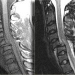
So I fall back on the routine of clearing the bones with a CT scan, and the ligaments with something else. That something else could be a clinical exam (not available in the obtunded patient), flexion-extension images under fluoroscopy (makes a lot of people nervous), keeping the patient in a collar for weeks (skin breakdown), or an MRI. The problem is that there is little guidance in the literature regarding how good MRI is or the best way to use it.

A recent paper in the Journal of Trauma retrospectively looked at 512 out of 17,000 patients (!) seen over 5 years at one trauma center who had both CT and MRI of the c-spine. They wanted to determine if MRI was of any value in cervical spine clearance. Only 150 met the inclusion criteria (GCS<13, no obvious neuro deficit, normal CT). Half of the MRIs were normal. Of the abnormal ones, 81% showed a ligamentous or soft tissue injury. None were deemed unstable and no specific management was needed for any of the abnormal scans.

The authors interpreted their data as showing that MRI provided no additional useful information. However, numbers were (very) small, so the likelihood of them seeing someone with an unstable ligamentous injury was low. Could it be that they showed that MRI detected stable injuries well, and that they could essentially remove the collar based on that?

Bottom line: We still don’t know how to use MRI for clearance. My bias (no good data I can find) is that it is good in suggesting ligamentous injury via nearby edema. If this injury involves only one set of ligaments, it is very likely a stable one and the collar can be removed. If it involves several groups of ligaments, that is probably not the case. And how soon do we have to get the MRI after injury? Some have suggested that 72 hours is the ideal window because edema decreases afterwards. Sounds reasonable, but I can’t find a shred of evidence in the literature. For now, I’ll get an MRI within 72 hours and if it is abnormal, pass the buck to my neurosurgical colleagues so they can gnash their teeth, too.

I would be very happy if someone can help me out and point me towards some good literature on this topic!

Reference: The value of cervical magnetic resonance imaging in the evaluation of the obtunded or comatose patient with cervical trauma, no other abnormal neurological findings, and a normal cervical computed tomography. J Trauma 72(3):699-702, 2012.

Why Do They Call Them Rounds?

Face it. Everyone uses this term. But where did it come from? After a little digging, I think I’ve found the answer. I’m sure someone will step forward and offer another explanation, but the origins of some of our traditions grow foggy with time.

Supposedly, the term “rounds” was introduced by Sir William Osler, the famed physician, while he was at Johns Hopkins hospital in Baltimore. The original building was built in the 1880s and had a round dome. Osler and his trainees had to walk circular hallways to see their patients. I’ve not been there, but looking at the picture above, the corners of the building appear to be octagonal patient wards as well.

The term has stuck with us, and today just about every discipline from prehospital to rehab medicine use it! If anybody has another theory or correction, please let me know!

Reference: CIRCULAR HOSPITAL WARDS: PROFESSOR JOHN MARSHALL’S CONCEPT AND ITS EXPLORATION BY THE ARCHITECTURAL PROFESSION IN THE 1880s. Medical History 32:426-448, 1988.

Are Femoral Traction Splints Okay In Open Fractures?

Application of traction splints to the femur can be a bit tricky, mostly because of the various indications and contraindications. The company that makes the Hare traction splint gives the sole indication as a suspected femur fracture, and the sole contraindication as an open femur fracture. In my mind, this is a bit too simplistic.

I agree that the traction splint should only be applied on femur fractures, known or suspected. However, there are a few more contraindications:

  • The patient should not have a posterior pelvic fracture. Unfortunately, prehospital providers don’t have xray vision, so they usually can’t tell. If there is any suspicion (pelvic instability, deformity), then don’t use it.
  • The knee joint must be intact. Application of a traction splint across a bad knee will distract the tibia and the femur, potentially causing more injury. Take a good look at the knee. If it’s edematous or discolored, no traction splint.
  • The tibia must not be fractured. As in the previous bullet point, the tibial segments will pull apart before the strong muscles in the thigh allow the femur to reduce.

What about the open fracture scenario? The concern is that contaminated bone will be pulled back into the wound. It’s not really known whether this results in an increased infection rate, but it’s better to be safe and not do it. However, there are two scenarios when applying traction to an open femur fracture is warranted:

  • There is significant bleeding from the wound. Restoring the normal anatomy will create more pressure around the injured tissues and may slow bleeding.
  • The distal pulses are compromised or absent. Most of the time, this is due to kinking of the vessel, not outright damage to it. Pulling it to length may restore normal flow.

Bottom line: Treat traction splints with respect. Keep these tips in mind, but always adhere to your local protocols and procedures first. However, if it’s not covered by them, or you are getting concerned that the patient’s (or their leg’s) wellbeing is at risk, do the right thing!

Thanks to Don Dustin from Mineral County EMS in Colorado for posing this question!

Figure It Out! The Answer

Several readers emailed the correct answer yesterday. The picture is a child with a bicycle handlebar injury to the epigastrium. The plastic grip over the end of the handlebar has a small hole in the middle leaving the distinctive mark seen in the photo.

Children are more likely to sustain significant injuries from this mechanism because they have little muscle in their abdominal wall, so it can’t protect as well as it does in adults. Everything between the handlebar and the spine gets crushed together, frequently resulting in serious injury.

Possible injuries include:

  • Pancreatic injury / transection
  • Liver laceration (left lobe)
  • Duodenal injury / hematoma
  • Retrohepatic vena cava injury

I’ve listed them in what I believe to be the usual order. The literature varies a bit because there aren’t a lot of series published. In this case, the injury was a pancreatic transection.

Bottom line: Handlebar injuries in children (and to a lesser degree, adults) are a significant marker for serious abdominal injury. CT scan is mandatory to find the diagnosis. Proper management of a pancreatic injury is a good topic for a future post!