All posts by The Trauma Pro

How Common Is BCVI?

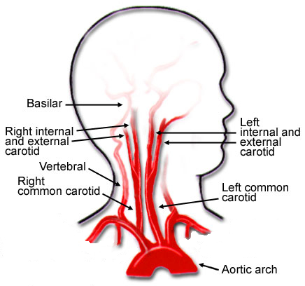
Blunt carotid and vertebral artery injuries (BCVI) are an under-appreciated problem after blunt trauma. Several screening tools have been published over the years, but they tend to be unevenly applied at individual trauma centers. I will discuss them in detail in the next section.

For the longest time, the overall incidence of BCVI was thought to be low, on the order of 1-2%. This is the number I learned years ago, and it has not really changed over time.

But how do we know for sure? Well, the group at Birmingham retrospectively reviewed every CT angiogram (CTA) of the neck they did in a recent two-year period. They did this after adopting a policy of imaging each and every one of their major blunt trauma patients for BCVI. Each patient chart was also evaluated to see if the patient met any of the criteria for the three commonly used screening systems.

During the study period, a total of 6,287 of 6,800 blunt trauma patients underwent BCVI screening with CTA of the neck. They discovered that 480 patients (7.6%) were positive for BCVI!

This is a shocking 8x higher than we expected! So why hasn’t this been obvious until now? Most likely because we were previously only aware of patients who became symptomatic. Luckily, many of these patients dodge the proverbial bullet and never exhibit any symptoms at all.

And what about pediatric patients? The neurosurgery groups at the University of New Mexico and Texas Children’s Hospital analyzed data in the Kids’ Inpatient Database (KID), which contains nationally representative pediatric data from the US. Five samples were obtained three years apart, beginning in 2000 and extending to 2012.

There were nearly 650,000 admissions for blunt trauma in the database, and 2150 were associated with BCVI. There was an interesting trend: incidence in 2000 started at 0.24% and increased to 0.49% in 2012. This represents a relative doubling of cases! Keep in mind that the absolute numbers remained very small, especially compared to the adult incidence.

Children aged 4 to 13 had the lowest risk of sustaining BCVI. This was higher in younger kids (ages 0-3), probably due to their big heads. It was also higher in adolescents and young adults (age 15-20). The injury was found more often in conjunction with cervical spine, skull base, clavicle, and facial fractures, as well as in children with TBI and intracranial hemorrhage.

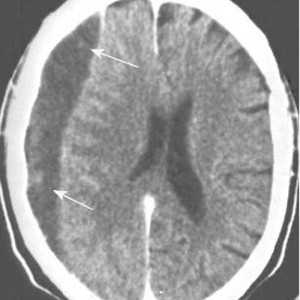
Over one-third of children sustaining BCVI suffered a stroke (37%). Mortality was high, with a total mortality of 13%. This increased to a 20% rate if a stroke occurred.

So why should we be worried? This is one of those clinical entities like blunt thoracic aortic disruption that potentially has terrible consequences if ignored. And it seems to be worse among children even though it is far less common. Although the number of patients who develop sequelae from their BCVI is small, suffering a stroke can be catastrophic.

Should we perform a screening study for all blunt trauma patients? It seems like overkill, or is it? Is there any way we can be more selective about it?

In the next post, I’ll review the current screening tools used to determine which patients should receive CTA and how good they are.

References:

  1. Universal screening for blunt cerebrovascular injury. J Trauma 90(2):224-231, 2021.
  2. Blunt cerebrovascular injury in pediatric trauma: a national database study. J Neurosurg Pediatr. 2019

Blunt Carotid And Vertebral Artery Injury (BCVI): First Adults, And Now In Children!

I’ve written quite a bit about the challenges of diagnosing blunt carotid and vertebral artery injury (BCVI) in adults.  And now some papers on the potential danger to pediatric patients are beginning to surface.

I think it’s time to repost and update my series on BCVI. I’ll start with the basics, like nomenclature. I’ll then move on to how often we actually encounter it. This will include new information on younger patients.

I’ll dig into the various screening systems, and will include new information from some recent pediatric papers. The, I’ll finish up with how to grade it and suggested treatment routines based on grade.

Lets start with the acronym itself. There seems to be some confusion as to what BCVI actually stands for. Some people believe that it means blunt cerebrovascular injury. This is not correct, because that term refers to injury to just about any vessel inside the skull.

The correct interpretation is blunt carotid and vertebral artery injury. This term refers to any portion and any combination of injury to those two pairs of vessels, from where they arise on the great vessels, all the way up into the base of the skull. Here’s a nice diagram:

Note that we will be excluding the external carotid arteries from this discussion, since injuries to them do not have any impact on the brain. They can cause troublesome bleeding, though.

These arteries are relatively protected from harm during blunt trauma. But given enough energy, bad things can happen. Fortunately, injuries to these structures are not very common, but unfortunately many trauma professionals under-appreciate their frequency and severity.

In the next post, we’ll explore the incidence of this injury in both adults and children. Is it truly as uncommon as we think?

Subdural Hematomas and Hygromas Simplified

There’s a lot of confusion about subdural pathology after head trauma. All subdural collections are located under the dura, on the brain’s surface. In some way, they involve or can involve the bridging veins, which are somewhat fragile and get more so with age.

Head trauma causes a subdural hematoma by tearing some of these bridging veins. Notice how thick the dura is and how delicate the bridging veins are in the image below.

When these veins tear, bleeding ensues, which layers out over the surface of the brain in that area. If the bleeding does not stop, pressure builds and compresses and shifts the brain. A subdural hematoma is considered acute from the time of injury until about three days later. During this time, it appears more dense than brain tissue.

After about 3-7 days, the clot begins to liquefy and becomes less dense on CT. Many hematomas are reabsorbed, but occasionally there is repeated bleeding from the bridging veins, or the hematoma draws fluid into itself due to the concentration gradient. As a result, it can enlarge and begin to cause new symptoms. During this period, it is considered subacute.

It moves on to a more chronic stage over the ensuing weeks. The blood cells in it break down completely, and the fluid that is left is generally less dense than the brain underneath it. The image below shows a chronic subdural (arrows).

Hygromas are different because they are a collection of CSF, not blood. They are caused by a tear in the meninges and allow CSF to accumulate in the subdural space. This can also be caused by head trauma and is generally very slow to form. They can lead to slow neurologic deterioration and are often found on head CT in patients with a history of falls, sometimes in the distant past. CT appearance is similar to a chronic subdural, but the density is the same as CSF, so it should have the same appearance as the fluid in the ventricle on CT.

The January Trauma MedEd Newsletter Is Available!

The January issue of the Trauma MedEd newsletter is now available to everyone!

This issue contains a collection of miscellaneous interesting stuff.

In this issue, you will learn about:

  • Nonsurgical Admissions And The Nelson Score
  • Tip: Evaluation of Hematuria in Blunt Trauma
  • Central Lines Cause Hypercoagulability?
  • Lab Values From Intraosseous Blood
  • Leukocytosis After Splenic Injury

To download the current issue, just click here! 

Or copy this link into your browser:  https://www.traumameded.com/courses/interesting-stuff-1/

This newsletter was released to subscribers a week ago. If you would like to be the first to get your hands on future newsletters, just click here to subscribe!

Does The Color Of Your Scrubs Matter?

In most hospitals, it seems that workers in every department wear a different color of scrubs. Traditionally, surgeons have worn scrubs in darker shades of green or blue. This is not always true, as some hospitals have adopted crazy colors in order to reduce theft. Apparently, not too many people are comfortable wearing a pilfered pair of bright pink scrubs in public.

We know that color can have subliminal impacts on people. Blue tends to have a calming effect. This is one of the reasons that police officer uniforms are frequently this color. Green and blue also tend to be associated with medicine. Red, orange,  and yellow are often associated with food. Ever wonder why McDonald’s arches are the color they are?

But what about scrubs? Patients do tend to form associations between a clinician’s dress and their intelligence, empathy, and trustworthiness. Interestingly, scrubs (as opposed to dress clothes) score high for all of these.

But what about the rainbow of colors that scrubs are available in? A recent research letter was submitted to JAMA Surgery by a group at UNC Chapel Hill. They administered an electronic survey over a two-month period to adult patients and visitors at their university hospital.  Their goal was to determine whether scrub color influenced the perception that the wearer was a surgeon, and what character traits were perceived.

This is a copy of the survey, asking for the identification of the surgeon, and the most skilled individual based on scrub color.

The results were quite interesting. This is a chart of trait identification based on color for men. The chart for women was very similar. Note that taller bars are a negative.

Here are the factoids:

  • Half of participants were 30-60 years old, and the remainder were evenly split between younger and older people
  • Green was the color most associated with surgeons and was selected by nearly half of participants. Sex did not seem to matter.
  • Black had the most negative connotation of any color
  • Blue scrubs were associated with the most caring clinicians; however it also implied that they were less knowledgeable, less skilled, and less trustworthy

Bottom line: This is an intriguing little study that shows that unfortunately, looks do matter. Even the colors of our clothes do! The participants associated black with death and said they looked like a mortician’s uniform. So definitely avoid!

The poor perception of clinicians wearing green scrubs is difficult to explain, but consistent. The navy and blue characteristics were generally positive and don’t look appreciably different from each other.

Hospitals pay little attention to the color of the scrubs they purchase. But this choice may have an impact on how the wearer is perceived by patients and families. Perhaps it is time to rethink color in patient-facing clinicians. And avoid black scrubs like the plague!

Reference: Association Between Patient Perception of Surgeons
and Color of Scrub Attire. JAMA Surg, 2023 Jan 11. doi: 10.1001/jamasurg.2022.5837. Epub ahead of print. PMID: 36630142.